1.1.1.379: (R)-mandelate dehydrogenase
This is an abbreviated version!
For detailed information about (R)-mandelate dehydrogenase, go to the full flat file.
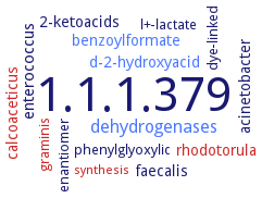
Word Map on EC 1.1.1.379
-
1.1.1.379
-
dehydrogenases
-
enterococcus
-
acinetobacter
-
2-ketoacids
-
calcoaceticus
-
faecalis
-
rhodotorula
-
benzoylformate
-
d-2-hydroxyacid
-
phenylglyoxylic
-
enantiomer
-
graminis
-
l+-lactate
-
dye-linked
-
synthesis
- 1.1.1.379
- dehydrogenases
-
enterococcus
-
acinetobacter
-
2-ketoacids
- calcoaceticus
-
faecalis
- rhodotorula
- benzoylformate
- d-2-hydroxyacid
-
phenylglyoxylic
-
enantiomer
- graminis
-
l+-lactate
-
dye-linked
- synthesis
Reaction
Synonyms
D(-)-mandelate dehydrogenase, D-mandelate dehydrogenase, D-ManDH, D-ManDH2, DMDH, LhDMDH, ManDH2, NAD+-dependent D-mandelate dehydrogenase, NAD-dependent D-mandelate dehydrogenase
ECTree

Advanced search results
Crystallization
Crystallization on EC 1.1.1.379 - (R)-mandelate dehydrogenase
Please wait a moment until all data is loaded. This message will disappear when all data is loaded.
 top
topCRYSTALLIZATION (Commentary) 

ORGANISM
UNIPROT
LITERATURE
purified enzyme in complex with substrates NADH and anilino(oxo)acetate (AOA), hanging-drop vapor diffusion method, mixing of 10 mg/ml protein in 5 mM sodium MOPS, pH 7.5, 2 mM NADH, and 20 mM AOA with reservoir solution containing 0.1 M cacodylate-Na, pH 5.0, 0.17 M ammonium sulfate, and 22.5% PEG 8000, in a 1:1 ratio, at 25°C, X-ray diffraction structure determination and analysis at 2.4 A resolution, molecular replacement using the crystal structure of unliganded D-ManDH2 (PDB ID 3WFI) as a search model, modeling. The ternary complex of D-ManDH2 forms a 2fold symmetric homodimer in a crystallographic asymmetric unit, as in the cases of the apo and binary complex structures
-
the enzyme is crystallized in three different forms using the hanging drop vapour diffusion method at 15-20°C. Type I crystals belong to space group P222(1), P22(1)2(1) or P2(1)2(1)2(1) with a = 100.3 A, b = 117.4 A, c = 80.4 A and are likely to contain a dimer in the crystallographic asymmetric unit