1.14.14.21: dibenzothiophene monooxygenase
This is an abbreviated version!
For detailed information about dibenzothiophene monooxygenase, go to the full flat file.

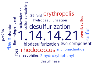
Word Map on EC 1.14.14.21 
-
1.14.14.21
-
desulfurization
-
rhodococcus
-
erythropolis
-
sulfur
-
flavin
-
biodesulfurization
-
two-component
-
2-hydroxybiphenyl
-
fossil
-
mononucleotide
-
sulfoxidation
-
paenibacillus
-
pet28a
-
dszabc
-
hydrodesulfurization
-
desulfinase
-
39-fold
-
sulfur-containing
-
flavin-dependent
-
mesophiles
- 1.14.14.21
-
desulfurization
- rhodococcus
- erythropolis
- sulfur
- flavin
-
biodesulfurization
-
two-component
- 2-hydroxybiphenyl
-
fossil
- mononucleotide
-
sulfoxidation
- paenibacillus
-
pet28a
-
dszabc
-
hydrodesulfurization
- desulfinase
-
39-fold
-
sulfur-containing
-
flavin-dependent
-
mesophiles
Reaction
Synonyms
BdsC, benzothiophene monooxygenase, BT monooxygenase, cofactor-requiring dibenzothiophene monooxygenase, DBT monooxygenase, DBT-MO, DBT-monooxygenase, dibenzothiophene monooxygenase, dszC, TdsC
ECTree
Advanced search results
Crystallization
Crystallization on EC 1.14.14.21 - dibenzothiophene monooxygenase
Please wait a moment until all data is loaded. This message will disappear when all data is loaded.
purified enzyme DszC, hanging drop vapour diffusion method, using a reservoir suliton containing 30% polyethylene glycol, 0.1 M sodium citrate, pH 5.6, and 0.2 M ammonium acetate, 4°C, X-ray diffraction structure determination and analysis
purified recombinant His-taged enzyme, hanging drop vapor diffusion method, mixing of 0.002 ml of 20 mg/ml protein in 20 mM Tris-HCl (pH 8.0) and 150 mM NaCl, with FMN in a 1:10 molar ratio, with 0.002 ml of reservoir solution containing 200 mM lithium sulfate, 100 mM Bis-Tris, pH 6.5, and 35% w/v PEG 3350, equilibration against 0.3 ml reservoir solution, 4°C, X-ray diffraction structure determination and analysis at 2.26 A resolution, molecular replacement using DszC structure, PDB ID 4JEK, as the search model
-
purified recombinant His-tagged enzyme in apoform and as FMN-bound enzyme DszC, two distinct conformations occur in the loop region (residues 131-142) adjacent to the active site, sitting drop vapor diffusion method, mixing of 0.001 ml protein solution with 0.001 ml of reservoir solution consisting of 0.2 M malonate, pH 6.0, 24% w/v PEG 3350, and 50 mM NaF, at 20°C, with or without 1 mM FMN, X-ray diffraction structure determination and analysis at 2.11 A an 2.3 A resolution, respectively, Each crystal contains two tetramers in the asymmetric unit, formed by two homodimers
purified recombinant wild-type and selenomethionine-labeled enzymes, hanging-drop vapour-diffusion method, mixing of 10 mg/ml wild-type enzyme in 20 mM Tris-HCl, pH 8.0, 150 mM NaCl with reservoir solution containing 17.5% PEG 3350, 200 mM Bis-Tris, pH 7.5, and 200 mM ammonium sulfate, and mixing of 5 mg/ml selenomethionine-labeled enzyme in 20 mM Tris-HCl, pH 8.0, 150 mM NaCl with reservoir solution containing 25% PEG 3350, 100 mM PIPES, pH 7.0, 200 mM ammonium sulfate, 20°C, X-ray diffraction structure determination and analysis at 2.4-2.9 A resolution
purified recombinant wild-type, selenomethionine-labeled, and mutant enzymes, hanging drop vapor diffusion method, mixing of 15-20 mg/ml protein in 10 mM Tris-HCl, pH 8.0, and 100 mM NaCl with an equal volume of reservoir solution containing 0.2 M L2SO4, 0.1 M Bis-Tris, pH 6.5, and 23% w/v PEG 3350, at 20°C, X-ray diffraction structure determination and analysis at 1.79 A resolution, molecular replacement and modelling


 results (
 results (
 top
top


