1.14.14.24: vitamin D 25-hydroxylase
This is an abbreviated version!
For detailed information about vitamin D 25-hydroxylase, go to the full flat file.

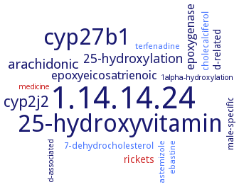
Word Map on EC 1.14.14.24 
-
1.14.14.24
-
25-hydroxyvitamin
-
cyp27b1
-
cyp2j2
-
arachidonic
-
25-hydroxylation
-
epoxyeicosatrienoic
-
epoxygenase
-
rickets
-
d-related
-
male-specific
-
7-dehydrocholesterol
-
cholecalciferol
-
astemizole
-
ebastine
-
terfenadine
-
d-associated
-
medicine
-
1alpha-hydroxylation
- 1.14.14.24
-
25-hydroxyvitamin
- cyp27b1
- cyp2j2
-
arachidonic
-
25-hydroxylation
-
epoxyeicosatrienoic
- epoxygenase
- rickets
-
d-related
-
male-specific
- 7-dehydrocholesterol
- cholecalciferol
- astemizole
- ebastine
- terfenadine
-
d-associated
- medicine
-
1alpha-hydroxylation
Reaction
Synonyms
CYP2R1, CYP24A1, CYP2C11, CYP2D25, CYP2J3, CYP2R1, CYPIIJ3, Cytochrome P450 2C11, cytochrome P450 2J2, Cytochrome P450 2J3, cytochrome P450 2R1, EC 1.14.13.159, VDH, vitamin D 25-hydroxylase, vitamin D-25-hydroxylase, vitamin D2 25-hydroxylase, vitamin D3 25-hydroxylase, vitamin D3 hydroxylase
ECTree
Advanced search results
Crystallization
Crystallization on EC 1.14.14.24 - vitamin D 25-hydroxylase
Please wait a moment until all data is loaded. This message will disappear when all data is loaded.
homology modeling. The relative position of Val391 in the beta3a-strand of a homology model and the crystal structure of rat CYP24A1 are consistent with hydrophobic contact of Val391 and the substrate side chain near C21
in complex with vitamin D3, to 1.0 A resolution. The CYP2R1 structure adopts a closed conformation with the substrate access channel being covered by the ordered B'-helix and slightly opened to the surface, which defines the substrate entrance point. The active site is lined by conserved, mostly hydrophobic residues. Vitamin D3 is bound in an elongated conformation with the aliphatic side-chain pointing toward the heme. The structure reveals the secosteroid binding mode in an extended active site


 results (
 results (
 top
top


