1.14.99.1: prostaglandin-endoperoxide synthase
This is an abbreviated version!
For detailed information about prostaglandin-endoperoxide synthase, go to the full flat file.
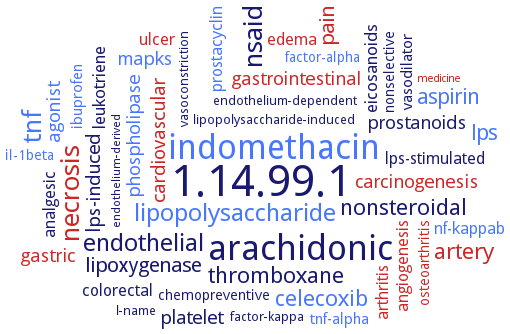
Word Map on EC 1.14.99.1
-
1.14.99.1
-
arachidonic
-
indomethacin
-
necrosis
-
nsaid
-
tnf
-
lipopolysaccharide
-
endothelial
-
nonsteroidal
-
celecoxib
-
artery
-
thromboxane
-
aspirin
-
lps
-
lipoxygenase
-
pain
-
cardiovascular
-
lps-induced
-
platelet
-
prostanoids
-
agonist
-
gastrointestinal
-
mapks
-
phospholipase
-
gastric
-
carcinogenesis
-
eicosanoids
-
leukotriene
-
ulcer
-
arthritis
-
analgesic
-
prostacyclin
-
nf-kappab
-
lps-stimulated
-
colorectal
-
angiogenesis
-
edema
-
tnf-alpha
-
ibuprofen
-
vasodilator
-
il-1beta
-
osteoarthritis
-
nonselective
-
factor-alpha
-
chemopreventive
-
lipopolysaccharide-induced
-
factor-kappa
-
endothelium-dependent
-
vasoconstriction
-
l-name
-
endothelium-derived
-
medicine
- 1.14.99.1
-
arachidonic
- indomethacin
- necrosis
-
nsaid
- tnf
- lipopolysaccharide
- endothelial
-
nonsteroidal
- celecoxib
- artery
-
thromboxane
- aspirin
- lps
- lipoxygenase
- pain
- cardiovascular
-
lps-induced
- platelet
-
prostanoids
- agonist
- gastrointestinal
- mapks
- phospholipase
- gastric
- carcinogenesis
-
eicosanoids
-
leukotriene
- ulcer
- arthritis
-
analgesic
- prostacyclin
- nf-kappab
-
lps-stimulated
- colorectal
- angiogenesis
- edema
- tnf-alpha
- ibuprofen
-
vasodilator
- il-1beta
- osteoarthritis
-
nonselective
- factor-alpha
-
chemopreventive
-
lipopolysaccharide-induced
-
factor-kappa
-
endothelium-dependent
-
vasoconstriction
-
l-name
-
endothelium-derived
- medicine
Reaction
Synonyms
(PG)H synthase, COX, COX-1, COX-2, COX1, Cox2, cyclooxygenase, cyclooxygenase 1, cyclooxygenase 2, cyclooxygenase-1, cyclooxygenase-1b, cyclooxygenase-2, cycloxigenase-2, fatty acid cyclooxygenase, hPGHS-1, hPGHS-2, oPGHS-1, PG G/H synthase 2, PG H synthase, PG synthetase, PG-endoperoxide synthase 2, PG-endoperoxide synthetase, PGH-synthase, PGHS, PGHS isoform-1, PGHS-1, PGHS-2, PHS, PHS-1, PHS-2, prostagladin-H synthase, prostaglandin endoperoxide H synthase, prostaglandin endoperoxide H synthase 1, prostaglandin endoperoxide H synthase 2, prostaglandin endoperoxide H synthase-1, prostaglandin endoperoxide H2 synthase-2, prostaglandin endoperoxide synthase, prostaglandin endoperoxide synthase 2, prostaglandin endoperoxide synthase-1, prostaglandin endoperoxide synthase-2, prostaglandin endoperoxide synthetase, prostaglandin G/H synthase, prostaglandin G/H synthase-2, prostaglandin H synthase, prostaglandin H synthase-1, prostaglandin H synthase-2, prostaglandin H2 synthase, prostaglandin H2 synthase-1, prostaglandin synthase, prostaglandin synthase-2, prostaglandin synthetase, prostaglandin-endoperoxide synthase, prostaglandin-endoperoxide synthase 1, prostaglandin-endoperoxide synthase 2, prostaglandin-H-synthase, prostaglandin-H-synthase 1, prostaglandin-H-synthase 2, PTGS 2, PTGS-1, PTGS-2, PTGS1, PTGS2, putative cyclooxygenase-3, synthase, prostaglandin, tPGHS-1, tPGHS-2
ECTree

Advanced search results
General Information
General Information on EC 1.14.99.1 - prostaglandin-endoperoxide synthase
Please wait a moment until all data is loaded. This message will disappear when all data is loaded.
 top
topGENERAL INFORMATION 

ORGANISM
UNIPROT
COMMENTARY 
LITERATURE
evolution
the algal PGHS lacks structural elements identified in all known animal PGHSs, such as epidermal growth factor-like domain and helix B in the membrane binding domain. The key residues of animal PGHS, like catalytic Tyr385 and heme liganding His388 are conserved in the algal enzyme, but the amino acid residues shown to be important for substrate binding and coordination, and the target residues for nonsteroidal anti-inflammatory drugs, Arg120, Tyr355, and Ser530, are not found at the appropriate positions in the algal sequences. The preferred substrate for the algal PGHS is arachidonic acid with cyclooxygenase reaction rate remarkably higher than values reported for mammalian PGHS isoforms
malfunction
metabolism
physiological function
additional information
malfunction

PGHS-1 inhibition in activated human plateletts significantly decreases PGHS-1-dependent thromboxane B2 formation in parallel with a decrease in platelet aggregation
malfunction
-
cervical distensibility is decreased in enzyme knockout mice on the day of expected delivery. Delayed parturition in enzyme knockout mice is the result of impaired luteolysis and cervical dilation
metabolism
-
the enzyme catalyzes the committed step in prostaglandin biosynthesis
metabolism
prostaglandin H synthase-1 catalyzes the first two steps in prostaglandin synthesis
metabolism
prostaglandin H synthase-2 catalyzes the first two steps in prostaglandin synthesis
physiological function
-
periovulatory expression of the Ptgs2 gene is essential for ovulation
physiological function
-
PGHS-2 also shows cyclooxygenase activity, which is implicated in colorectal cancer
physiological function
-
PHS isozyme-dependent oxidative damage to proteins and DNA, and cytotoxicity, overview. hPHS-1- and hPHS-2-expressing cells incubated with dopamine, L-dihydroxyphenylalanine, dihydroxyphenylacetic acid, or homovanillic acid exhibit increased cytotoxicity compared to untransfected cells, and cytotoxicity is increased further by exogenous arachidonic acid, which increases hPHS activity. Isozyme-specific, PHS-dependent oxidative damage and cytotoxicity caused by neurotransmitters, their precursors, and their metabolites may contribute to neurodegeneration associated with aging
physiological function
prostaglandin H synthases or cyclooxygenases catalyse the peroxidation of arachidonic acid to PGG2 and PGH2 which are further converted to a series of prostaglandins and thromboxane A2. The enzyme catalyzes formation of oxidative stress biomarkers malondialdehyde and 15(S)-8-iso-prostaglandin F2alpha, and other F2-isoprostanes, promoted by glutathione
physiological function
prostaglandin-endoperoxide synthases and nitric oxide synthases regulate the bovine corpea lutea life span mainly during the transition from the luteotrophic to the luteolytic phase
physiological function
PTGS2 plays a pivotal role in inflammation, tissue damage, and tumorigenesis. Differential regulation of PCSKs by PTGS2 with a pivotal role for E2F1, implication of PTGS2-derived PGE2 as a determining factor in the regulation of PCSK activity
physiological function
the bi-functional enzyme with cyclooxygenase and peroxidase activities plays a central role in the inflammatory response, pain, and blood clotting
physiological function
-
the enzyme promotes Col10a1 expression and enhances hypertrophic differentiation of ATDC5 cells. Cox-2 also upregulates marker genes of chondrocyte maturation, apoptosis, and matrix mineralization, including transcription factors Runx2, Alp, Bsp, and Osterix
additional information
-
aqueous extracts of Chromoleana odorata, commonly used in traditional medicine as antiinflammatory drug against pains or as cataplasm to stop hemorrhage in Ivory Coast, the essential oil extracted from the fresh leaves activates the cyclooxygenase activity of the PGHS, overview
additional information
-
neurotoxicity of the amphetamine analogues methamphetamine and 3,4-methylenedioxyamphetamine, the active metabolite of ecstasy, may involve their prostaglandin H synthase-dependent bioactivation to free radical intermediates that generate reactive oxygen species and oxidatively damage cellular macromolecules. The activation effect is blocked by irreversible enzyme inhibitor acetylsalicylic acid, overview
additional information
-
PHS is involved in the mechanism of thalidomide to cause increased embryonic DNA oxidation measured as 8-oxoguanine leading to embryopathies, phenotype, overview. A prostaglandin H synthase-dependent, reactive oxygen species-mediated mechanism. Thalidomide teratogenicity was blocked by maternal pretreatment with acetylsalicylic acid, an irreversible inhibitor of prostaglandin H synthase
additional information
seminal plasma-induced PTGS2 expression is mediated by intracellular signaling pathways involving MAPKs and NF-kappaB. Depending on cell type and stimulus, different intracellular signaling pathways are involved in inflammation and PTGS2 expression
additional information
-
seminal plasma-induced PTGS2 expression is mediated by intracellular signaling pathways involving MAPKs and NF-kappaB. Depending on cell type and stimulus, different intracellular signaling pathways are involved in inflammation and PTGS2 expression