2.2.1.1: transketolase
This is an abbreviated version!
For detailed information about transketolase, go to the full flat file.

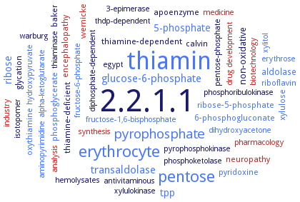
Word Map on EC 2.2.1.1 
-
2.2.1.1
-
thiamin
-
pentose
-
erythrocyte
-
pyrophosphate
-
transaldolase
-
glucose-6-phosphate
-
tpp
-
ribose
-
5-phosphate
-
aldolase
-
non-oxidative
-
glycation
-
encephalopathy
-
pyridoxine
-
apoenzyme
-
phosphoglycerate
-
wernicke
-
baker
-
oxythiamine
-
neuropathy
-
ribose-5-phosphate
-
thiamine-deficient
-
xylulose
-
thiamine-dependent
-
6-phosphogluconate
-
riboflavin
-
calvin
-
pharmacology
-
drug development
-
biotechnology
-
pentose-phosphate
-
xylulokinase
-
industry
-
alpha-ketoglutarate
-
dihydroxyacetone
-
warburg
-
phosphoketolase
-
3-epimerase
-
hemolysates
-
pyrophosphokinase
-
xylitol
-
phosphoribulokinase
-
thiaminase
-
hydroxypyruvate
-
aminopyrimidine
-
fructose-6-phosphate
-
medicine
-
fructose-1,6-bisphosphate
-
antivitaminous
-
erythrose
-
egypt
-
thdp-dependent
-
diphosphate-dependent
-
synthesis
-
isotopomer
-
analysis
- 2.2.1.1
- thiamin
- pentose
- erythrocyte
- pyrophosphate
- transaldolase
- glucose-6-phosphate
- tpp
- ribose
- 5-phosphate
- aldolase
-
non-oxidative
-
glycation
- encephalopathy
- pyridoxine
-
apoenzyme
- phosphoglycerate
- wernicke
-
baker
- oxythiamine
- neuropathy
- ribose-5-phosphate
-
thiamine-deficient
- xylulose
-
thiamine-dependent
- 6-phosphogluconate
- riboflavin
-
calvin
- pharmacology
- drug development
- biotechnology
-
pentose-phosphate
- xylulokinase
- industry
- alpha-ketoglutarate
- dihydroxyacetone
-
warburg
- phosphoketolase
-
3-epimerase
- hemolysates
-
pyrophosphokinase
- xylitol
- phosphoribulokinase
- thiaminase
- hydroxypyruvate
- aminopyrimidine
- fructose-6-phosphate
- medicine
- fructose-1,6-bisphosphate
-
antivitaminous
- erythrose
-
egypt
-
thdp-dependent
-
diphosphate-dependent
- synthesis
-
isotopomer
- analysis
Reaction
Synonyms
glycolaldehydetransferase, STM14_2885, STM14_2886, TK16, TKA, TKL, TKL1, Tkl2, TKT, TKT10, TKT3, TKT7, TktA, TktB, TKTc, TKTL-1, TKTL1, TKTL2, TKTp, transketolase, transketolase 10, transketolase 3, transketolase 7, transketolase A, transketolase B, transketolase like 1, transketolase-1, transketolase-like 1, transketolase-like enzyme 1, transketolase-like-1, transketolase-like-1-gene, transketolase-like-2
ECTree
Advanced search results
Crystallization
Crystallization on EC 2.2.1.1 - transketolase
Please wait a moment until all data is loaded. This message will disappear when all data is loaded.
apoenzyme and enzyme in complex with thiamine diphosphate and Mg2+, hanging drop vapor diffusion method, using 10% (w/v) PEG 6K, 5 % (v/v) 2-m,ethyl-2,4-pentanediol and 0.1 M MES pH 6.5-7.0 or 0.1 M HEPES pH 7.0-8.0
hanging drop vapor diffusion method, transketolase in a covalent complex with donor ketoses D-xylulose 5-phosphate and D-fructose 6-phosphate at 1.47 A and 1.65 A resolution, reveal significant strain in the tetrahedral cofactor-sugar adducts with a 25-30° out-of-plane distortion of the C2-Calpha bond connecting the carbonyl of the substrates with the C2 of the cofactors thiazolium part. The noncovalent complex with acceptor aldose ribose 5-phosphate reveals that the sugar is present in multiple forms, in a strained ring-closed beta-D-furanose form in C2-exo conformation as well as in an extended acyclic aldehyde form, with the reactive C1 aldo function held close to Calpha of the presumably planar carbanion/enamine intermediate
mutant S385Y/D469T/R520Q, hanging drop vapor diffusion method, using 17-22% (w/v) PEG 6000, 2% (v/v) glycerol, 50 mM glycyl-glycine buffer, pH 7.9
homology modeling of human transketolase using the crystal structure of yeast as a template, refinement of the model through molecular dynamics simulations. Five critical sites containing arginines R101, R318, R395, R401 and R474 contribute to dimer stability or catalytic activity. R101 and R401 maintain hydrogen bonds within the dimer, the most important being with D424 and E432, respectively. Both bonds are formed by charged residues. Non-conserved R395 also forms stable intermolecular hydrogen bonds. There is a substrate channel similar to the yeast enzyme
-
modeling of inhibitors 1-(3-chloro-2-methylphenyl)-3-(2-hydroxy-5-nitrophenyl)urea, 1-(5-hydroxynaphthalen-1-yl)-3-(2-methyl-5-nitrophenyl)urea, 1-(5-chloro-2-hydroxy-4-nitrophenyl)-3-phenylurea into enzyme structure model. the binding mode of the compounds involves interactions with the alpha helix sequence D200-G210 interfering likely with the enzyme dimerization
to 1.75 A resolution, space group C2 with one monomer in the asymmetric unit. Two monomers form the homodimeric biological assembly with two identical active sites at the dimer interface. The protomer exhibits the typical three alpha/beta-domain structure and topology reported for transketolases from other species, with structural differences for several loop regions and the linker that connects the diphosphate and pyridine domain. Two lysines and a serine interact with the beta-phosphate of thiamine diphosphate. Residue Gln189 spans over the thiazolium moiety of thiamine diphosphate and replaces an isoleucine found in most non-mammalian transketolases. The side chain of Gln428 forms a hydrogen bond with the 4-amino group of thiamine diphosphate and replaces a histidine that is invariant in all nonmammalian transketolases. All other amino acids involved in substrate binding and catalysis are strictly conserved. Formation of the central 1,2-dihydroxyethyl-thiamine diphosphate carbanion-enamine intermediate is thermodynamically favored with increasing carbon chain length of the donor ketose substrate
in complex with thiamine diphosphate, hanging drop vapor diffusion method, enzyme in complex with thiamine diphosphate using 15% (w/v) PEG 3350, 0.1 M sodium acetate, 0.1 M bis-Tris propane pH 7.5, apoprotein using 20% (w/v) PEG 3350, 0.2 M NaCl
to 2.3 A resolution. Crystals belong to trigonal space group P3221with unit-cell parameters a = b = 75.43, c = 184.11 A
to 2.5 A resolution. Final model includes 410 water molecules, four thiamine diphosphate molecules, 4 Mg2+ ions and eight glycerol molecules. The enzyme forms a homodimer, where the two monomeric units are related by a non-crystallographic twofold axis. There are two dimers per asymmetric unit that are related to each other by a twofold axis to create a noncrystallographic screw axis parallel to the crystallographic Z-axis
cocrystallization of apotransketolase with 5 mM thiamine diphosphate, 5 mM CaCl2, 50 mM fructose-6-phosphate, 13-16% (w/W) polyethylenglycol 6000 in 50 mM glycyl-glycine buffer, pH 7.6, 0.0075 ml of a 20 mg/ml solution mixed with the same amount of mother liquid, space group: P212121
wild-type and mutant E418A in complex with N1-methyl-thiamine diphosphate and wild-type in complex with 4-methylamino-thiamine diphosphate, topology of active sites remains unchanged by cofactor analogues
-
wild type and mutants H261F, H66C/261C, R356L and R525L enzymes in complex with various physiological ligands, hanging drop vapor diffusion method, using 0.1 M MES at pH 6.5, 0.1M NaCl, 30% (w/V) PEG 400


 results (
 results (
 top
top


