2.3.1.1: amino-acid N-acetyltransferase
This is an abbreviated version!
For detailed information about amino-acid N-acetyltransferase, go to the full flat file.
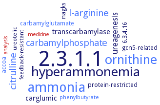
Word Map on EC 2.3.1.1
-
2.3.1.1
-
ammonia
-
hyperammonemia
-
ornithine
-
l-arginine
-
citrulline
-
carbamylphosphate
-
ureagenesis
-
transcarbamylase
-
carglumic
-
accoa
-
6.3.4.16
-
gcn5-related
-
protein-restricted
-
carbamylglutamate
-
nagks
-
feedback-resistant
-
phenylbutyrate
-
ureotelic
-
analysis
-
medicine
- 2.3.1.1
- ammonia
-
hyperammonemia
- ornithine
- l-arginine
- citrulline
- carbamylphosphate
-
ureagenesis
-
transcarbamylase
-
carglumic
- accoa
-
6.3.4.16
-
gcn5-related
-
protein-restricted
- carbamylglutamate
-
nagks
-
feedback-resistant
- phenylbutyrate
-
ureotelic
- analysis
- medicine
Reaction
Synonyms
acetylglutamate synthase, acetylglutamate synthetase, acetylglutamic synthetase, acetyltransferase, amino acid, AGAS, amino acid acetyltransferase, ARG2, ArgA, ArgH(A), argJ, Cg3035, More, N-acetyl-glutamate synthase, N-acetyl-L-glutamate synthase, N-acetyl-L-glutamate synthase/kinase, N-acetyl-L-glutamate synthetase, N-acetylglutamate synthase, N-acetylglutamate synthase/kinase, N-acetylglutamate synthetase, NAGS, NAGS-K, NAGS/K, NAT, ngNAGS, PaNAGS, pitax, Rv2747, SINAGS1, XcNAGS
ECTree

Advanced search results
Crystallization
Crystallization on EC 2.3.1.1 - amino-acid N-acetyltransferase
Please wait a moment until all data is loaded. This message will disappear when all data is loaded.
 top
topCRYSTALLIZATION (Commentary) 

ORGANISM
UNIPROT
LITERATURE
catalytic N-acetyltransferase domain complexed with N-acetyl-L-glutamate, hanging drop vapor diffusion method, using 100 mM Bis-Tris, pH 6.5, 35% (w/v) PEG3350
development of a structural model of human enzyme that is fully consistent with the functional effects of the 14 missense mutations that have been identified in N-acetylglutamate synthase-deficient patients
-
purified recombinant catalytic N-acetyltransferase domain complexed with N-acetyl-L-glutamate, sitting drop vapour diffusion method, mixing of 0.002 ml of 20 mg/ml protein in 50 mM Tris-HCl, pH 7.4, 50 mM NaCl, 10% glycerol, 5 mM bmercaptoethanol, and 1 mM EDTA, 10 m CoA, and 10 mM N-acetyl-L-glutamate, with 0.002 ml of reservoir solution containing 100 mM Bis-Tris, pH 6.5, 35% PEG 3350, 18°C, X-ray diffraction structure determination and analysis at 2.1 A resolution, molecular replacement
N-acetyl-L-glutamate synthase/kinase in complex with L-arginine, sitting drop vapor diffusion method, using 100 mM sodium cacodylate trihydrate, pH 6.2, 25% (w/v) polypropylene glycol P400 and 200 mM magnesium chloride
to 2.7 A resolution. Enzyme is a tetramer. Each subunit has an amino acid kinase domain, which is likely responsible for N-acetylglutamate kinase activity and has a putative arginine binding site, and an N-acetyltransferase domain that contains the putative N-acetylglutamate synthase active site. The angle of rotation between amino acid kinase and N-acetyltransferase domains varies among crystal forms and subunits within the tetramer. A rotation of 26° is sufficient to close the predicted AcCoA binding site, thus reducing enzymatic activity
in complex with with AcCoA and L-glutamate. The overall structure adopts a classic fold of the GCN5-related N-acetyltransferase family, characterized by a V-shaped cleft and beta-bulge. Activity depends on dimerization to form a deep, vast pocket for L-glutamate binding. L-glutamate binds at a site far away from AcCoA. A one-step sequential mechanism is proposed for enzymatic catalysis
structures complexed with CoA and product bound N-acetylglutamine and the other complexed with acetyl-CoA and the inhibitor L-arginine at 2.3 and 3.0 A resolution, respectively. The ArgA protomer has a V cleft and a beta bulge. ArgA may also acetylate L-glutamine like L-glutamate. The active site is strongly inhibited by L-arginine resulting in a closed conformation of ArgA. Both L-arginine and N-acetylglutamine occupy the same active site
hanging drop method
purified recombinant enzyme, hanging drop vapour diffusion method, mixing of 0.001 ml protein solution containing 13 mg/ml protein with 0.001 ml of reservoir solution containing 0.1 M Tris-HCl, pH 6.5, 1.6 M ammonium sulfate, and 0.1% w/v sodium azide, 18°C, 1-2 days, cryoprotection in 0.1 M Tris-HCl, pH 6.5, 1.6 M ammonium sulfate, 30% v/v glycerol and 0.1% w/v sodium azide, X-ray diffraction structure determination and analysis at 2.25 A resolution
sitting drop and hanging-drop vapor diffusion method, crystals belong to the hexagonal space group P6(2)22, with unit-cell parameters a = b = 134.60, c = 192.11 A, and diffract to about 3.0 A resolution
-
enzyme bound to N-acetyl-L-glutamate, sitting drop vapor diffusion method, using 0.2 M Li2SO4, 0.1 M Tris pH 6.5, 25% (w/v) PEG 3350
purified recombinant N-acetyltransferase domain of N-acetyl-L-glutamate synthase/kinase, with and without a His-tag, complexed with N-acetyl-L-glutamate, sitting-drop vapor-diffusion method, 0.002 ml of protein in 50 mM Tris-HCl, pH 7.4, 50 mM NaCl, 10% glycerol, 5 mM 2-mercaptoethanol, 1 mM EDTA, 10 mM CoA, and 10 mM N-acetyl-L-glutamate, is mixed with 0.2 M Li2SO4, 0.1 M Tris, pH 6.5, 25% PEG 3350 for the His-tagged enzyme, and with 0.2 M Li2SO4, 0.1 M Tris, pH 8.5, 25% PEG 3350 for the detagged enzyme, X-ray diffraction structure determination and analysis at 1.7 A and 1.4 A resolution, respectively