2.4.2.28: S-methyl-5'-thioadenosine phosphorylase
This is an abbreviated version!
For detailed information about S-methyl-5'-thioadenosine phosphorylase, go to the full flat file.
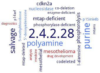
Word Map on EC 2.4.2.28
-
2.4.2.28
-
purine
-
polyamine
-
salvage
-
mtap-deficient
-
cdkn2a
-
mesothelioma
-
phosphorolysis
-
mesothelial
-
nucleosidase
-
codeleted
-
l-alanosine
-
co-deletion
-
mtap-deleted
-
phosphorylase-deficient
-
p15ink4b
-
5'-deoxyadenosine
-
5-methylthioribose
-
mtans
-
p14arf
-
enzyme-deficient
-
diagnostics
-
drug development
-
medicine
-
analysis
- 2.4.2.28
- purine
- polyamine
-
salvage
-
mtap-deficient
-
cdkn2a
- mesothelioma
-
phosphorolysis
-
mesothelial
- nucleosidase
-
codeleted
-
l-alanosine
-
co-deletion
-
mtap-deleted
-
phosphorylase-deficient
- p15ink4b
- 5'-deoxyadenosine
- 5-methylthioribose
-
mtans
-
p14arf
-
enzyme-deficient
- diagnostics
- drug development
- medicine
- analysis
Reaction
Synonyms
5'-deoxy-5'-methylthioadenosine phosphorylase, 5'-deoxy-5'-methylthioadenosine phosphorylase II, 5'-deoxy-5'-methylthioadenosine:orthophosphate methylthioribosyltransferase, 5'-methylthioadenosine nucleosidase, 5'-methylthioadenosine phosphorylase, 5'-methylthioadenosine/S-adenosylhomocysteine nucleosidase, 5-methylthioadenosine phosphorylase, 5�-methylthioadenosine phosphorylase, MeSAdo phosphorylase, MeSAdo/Ado phosphorylase, methylthioadenosine nucleosidase, methylthioadenosine nucleoside phosphorylase, methylthioadenosine phosphorylase, MTA phosphorylase, MTAN, MTAN1, MTAN2, MTAP, Mtap protein, MTAPase, MTN1, MTN2, PfMTAP, phosphorylase, methylthioadenosine, RSFP, Rv0535, SsMTAP, SsMTAP II, SsMTAPII, SSO2343
ECTree

Advanced search results
Crystallization
Crystallization on EC 2.4.2.28 - S-methyl-5'-thioadenosine phosphorylase
Please wait a moment until all data is loaded. This message will disappear when all data is loaded.
 top
topCRYSTALLIZATION (Commentary) 

ORGANISM
UNIPROT
LITERATURE
in complex with adenine, by sitting- and hanging-drop formation at 22°C, at 2.9 A resolution. Belongs to space group P3121, monomer consists of seven alpha-helices, ten beta-strands, and a 3(10)-helix. Residues between 216 and 225 demonstrate weak electron density for both subunits, therefore indicating this loop has high flexibility. Bound adenine is located in the deep pocket formed by the monomer with the entrance partially covered by the adjacent subunit. This flap is critical in the formation of a wide dimer interface
MTAN1 in its apo form and in complex with formycin A and 5'-methylthiotubercidin to a resolution of 2.0 A, 1.9 A and 1.8 A, respectively. By hanging-drop, vapor-diffusion technique. Belongs to space group P21. The monomer of MTAN1 has a mixed alpha/beta structure composed of a twisted, ten-stranded mixed beta sheet flanked by seven alpha helices and two short 3(10) helices. In the MTAN1 crystals, the asymmetric unit contains two monomers, which interact via an interface involving the four loops beta2-beta3, beta4-alpha2, beta6-alpha2b and beta8-alpha4, and the alpha2, alpha2b, and alpha5 helices. Both polar and apolar residues are involved in hydrogen bond and van der Waals interactions
in complex with adenine, hanging drop vapor diffusion method, using 20% (w/v) PEG-1000, 0.1 M phosphate-citrate (pH 4.2) and 0.2 M Li2SO4
the intersubunit interface of MTAN is highly hydrophobic and is dominated by van der Waals and hydrophobic interactions
-
crystal structures of apo MTAP and MTAP in complex with p-Cl-PhT-DADMe-ImmA are determined at 1.9 and 2.0 A resolution, respectively. Inhibitor binding causes condensation of the enzyme active site, reorganization at the trimer interfaces, the release of water from the active sites and subunit interfaces, and compaction of the trimeric structure. These structural changes cause the entropy-favored binding of transition state analogues
from recombinant enzyme, hanging drop-vapour diffusion method, protein 25 mg/ml, 10 mM Tris-HCl, pH 7.4, 5 mM DTT, 0.4 M NaCl against reservoir solution of 80 mM MES, pH 5.9, 5 mM DTT, 12% w/v polyethylene glycol 6000, 25% v/v ethylene glycol, in presence of substrates adenine, phosphate and sulfate, structure analysis by X-ray diffraction at light scattering
-
in complex with 5'-methylthio-DADMe-immucillin A, 5'-butylthio-DADMe-immucillin A, 5'-propylthio-immucillin A or 5'-methylthio-tubercidin, sitting drop vapor diffusion method, using 0.2 M magnesium chloride, 0.1 M sodium citrate:citric acid (pH 5.5), and 40% (w/v) PEG 400 for 5'-methylthio-DADMe-immucillin A or 5'-propylthio-immucillin A, 3 M sodium chloride and 0.1 M sodium acetate (pH 4.5) for BT-DADMe-immucillin A, and 0.2 M magnesium chloride and 20% (w/v) PEG 3350 for 5'-methylthio-tubercidin
-
X-ray structure with (1S)-1-(9-deazaadenin-9-yl)-1,4-di-deoxy-1,4-imino-5-methylthio-D-ribitol
analysis of the crystal packing. Space group is C2, unit-cell parameters are a = 135.16, b = 138.09, c = 96.56 A, beta = 92.21°. The asymmetric unit contains two independent half-hexamers, the other half of each of which is generated by a crystallographic twofold axis
crystals are grown at either room temperature or 18°C using the hanging drop vapor diffusion technique. Determination of the structure of 5'-deoxy-5'-methylthioadenosine phosphorylase alone, as ternary complexes with sulfate plus substrates 5'-deoxy-5'-methylthioadenosine, adenosine, or guanosine, or with the noncleavable substrate analog formycin B and as binary complexes with phosphate or sulfate alone. The structure of unliganded SsMTAP is refined at 2.5 A resolution and the structures of the complexes are refined at resolutions ranging from 1.6 A to 2.0 A
from recombinant enzyme, hanging drop-vapour diffusion method, protein solution, 7-10 mg/ml, 18°C, reservoir solution for native crystals: Tris-HCl 10 mM, pH 7.4, 28-30% dioxane, 12% 2-methyl-2,4-pentanediol, 0.12 M MgCl2, 0,04 M NaCl, for crystals of enzyme complexed with substrates or sulfate and phosphate ions, substrates are added and NaCl is exchanged for MgSO4 or NH4Cl and KH2PO4, respectively, X-ray structure determination and analysis
-
hanging-drop, vapor-diffusion method at 22°C, the crystal structure of the enzyme in complex with 5'-deoxy-5'-methylthioadenosine and sulfate is determined to 1.45 A resolution
the structure of SsMTAP II is originally determined in space group P1 and shows R32 pseudosymmetry. Post-analysis using phenix.xtriage shows that the correct space group is C2. The structure refined in space group C2 is reported and the factors that initially led to the incorrect space-group assignment are discussed
in complex with adenine, 5'-deoxy-5'-methylthioadenosine, tubercidin or sulfate, hanging drop vapor diffusion method, using 100 mM Bis-Tris or MES buffer, pH 6.1-6.7 and 14-18% (w/v) PEG 3350
apoenzyme and in complex with rhodamine B, hanging drop vapor diffusion method, using 35% (w/v) PPG 400, 75 mM MgCl2 and 0.1 M Bis-Tris pH 6.5