2.5.1.48: cystathionine gamma-synthase
This is an abbreviated version!
For detailed information about cystathionine gamma-synthase, go to the full flat file.
Word Map on EC 2.5.1.48
-
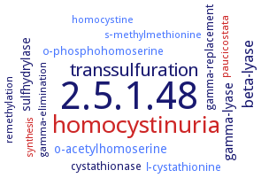
2.5.1.48
-
homocystinuria
-
transsulfuration
-
beta-lyase
-
gamma-lyase
-
sulfhydrylase
-
o-acetylhomoserine
-
gamma-replacement
-
l-cystathionine
-
cystathionase
-
o-phosphohomoserine
-
paucicostata
-
gamma-elimination
-
homocystine
-
s-methylmethionine
-
remethylation
-
synthesis
- 2.5.1.48
- homocystinuria
-
transsulfuration
-
beta-lyase
-
gamma-lyase
-
sulfhydrylase
- o-acetylhomoserine
-
gamma-replacement
- l-cystathionine
- cystathionase
- o-phosphohomoserine
- paucicostata
-
gamma-elimination
- homocystine
- s-methylmethionine
-
remethylation
- synthesis
Reaction
Synonyms
AtCGS, CGS, CGS1, CS,26, cystathionine gamma-synthase, cystathionine synthase, cystathionine synthetase, cystathionine-gamma-synthase, EC 4.2.99.9, homoserine O-transsuccinylase, homoserine transsuccinylase, HTS, MetB, O-succinyl-L-homoserine succinate-lyase (adding cysteine), O-succinylhomoserine (Thiol)-lyase, O-succinylhomoserine synthase, O-succinylhomoserine synthetase, synthase, cystathionine gamma-
ECTree

Advanced search results
Crystallization
Crystallization on EC 2.5.1.48 - cystathionine gamma-synthase
Please wait a moment until all data is loaded. This message will disappear when all data is loaded.
 top
topCRYSTALLIZATION (Commentary) 

ORGANISM
UNIPROT
LITERATURE
crystal structure at 1.5 A resolution. The pyridoxal phosphate cofactor is covalently bound to Lys204 via a Schiff base linkage in the deep cavity. Thr347 from the beta10-beta11 connecting loop, located at the entrance of the active site, is speculated to be a main contributor for stabilization of the acetyl group of O-acetyl-L-homoserine. Structural comparison with the structures of MetB from Nicotiana tabacum and Escherichia coli indicates that the conformation of the beta10-beta11 connecting loops determines the size and shape of the acetyl- or succinyl-group binding site and ultimately determines the substrate specificity
in silico modeling and pyridoxal 5'-phosphate cofactor docking study
-
to 1.9 A resolution. Cofactor pyridoxal 5'-phosphate binds tightly to Lys208 with a covalent-bond length ranging between 1.3 and 1.4 A. The cofactor is stabilized by a series of hydrogen bonds from Gly86, Met87, Asn158, Asp183 and Ser205 from one monomer and Tyr56 and Arg58 from the second monomer
crystals grown by sitting drop vapour diffusion against a reservoir containing 100 mM MES-NaOH
-
hanging drop vapor diffusion method, using either 100 mM acetate pH 3.6, 54% (w/v) 2-methyl-2,4-pentanediol, 200 mM magnesium chloride and 10 mM ammonium sulfate or 100 mM Tris-HCl pH 7.0, 12-18% (w/v) polyethylene glycol 3350, 200 mM sodium citrate and nickel(II) chloride hexahydrate
-
three crystal forms from different temperature and pH conditions, collected to 2.2, 2.9 and 2.7 A resolution for forms I, II and II', respectively. Form I crystals (space group P21, unit-cell parameters a = 58.4, b = 149.3, c = 90.2 A, beta = 108.9°) are obtained at 20°C under acidic pH conditions using 2-methyl-2,4-pentanediol. Under basic pH conditions the enzyme crystallizes in form II at 20°C(space group C2221, unit-cell parameters a = 117.7, b = 117.8, c = 251.3 A) and in form II' at 40°C (space group C2221, unit-cell parameters a = 107.5, b = 127.7, c = 251.1 A) using polyethylene glycol 3350
-