2.7.1.20: adenosine kinase
This is an abbreviated version!
For detailed information about adenosine kinase, go to the full flat file.

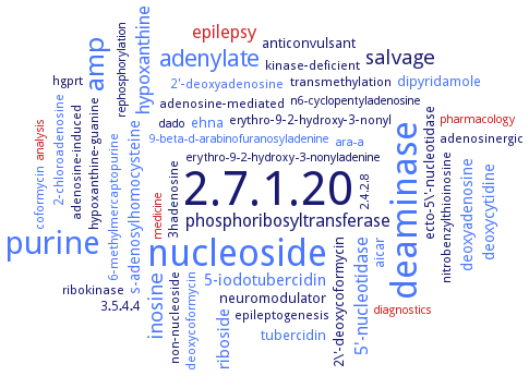
Word Map on EC 2.7.1.20 
-
2.7.1.20
-
nucleoside
-
deaminase
-
purine
-
amp
-
adenylate
-
inosine
-
salvage
-
hypoxanthine
-
epilepsy
-
phosphoribosyltransferase
-
5'-nucleotidase
-
5-iodotubercidin
-
riboside
-
deoxyadenosine
-
s-adenosylhomocysteine
-
deoxycytidine
-
aicar
-
anticonvulsant
-
tubercidin
-
dipyridamole
-
neuromodulator
-
ehna
-
2\'-deoxycoformycin
-
ecto-5\'-nucleotidase
-
ribokinase
-
erythro-9-2-hydroxy-3-nonyl
-
adenosinergic
-
6-methylmercaptopurine
-
3hadenosine
-
2-chloroadenosine
-
coformycin
-
nitrobenzylthioinosine
-
epileptogenesis
-
hypoxanthine-guanine
-
transmethylation
-
adenosine-induced
-
hgprt
-
kinase-deficient
-
2'-deoxyadenosine
-
adenosine-mediated
-
3.5.4.4
-
deoxycoformycin
-
non-nucleoside
-
analysis
-
2.4.2.8
-
9-beta-d-arabinofuranosyladenine
-
n6-cyclopentyladenosine
-
erythro-9-2-hydroxy-3-nonyladenine
-
dado
-
ara-a
-
medicine
-
diagnostics
-
rephosphorylation
-
pharmacology
- 2.7.1.20
- nucleoside
- deaminase
- purine
- amp
- adenylate
- inosine
-
salvage
- hypoxanthine
- epilepsy
-
phosphoribosyltransferase
- 5'-nucleotidase
- 5-iodotubercidin
- riboside
- deoxyadenosine
- s-adenosylhomocysteine
- deoxycytidine
- aicar
-
anticonvulsant
- tubercidin
- dipyridamole
-
neuromodulator
- ehna
-
2\'-deoxycoformycin
-
ecto-5\'-nucleotidase
- ribokinase
-
erythro-9-2-hydroxy-3-nonyl
-
adenosinergic
- 6-methylmercaptopurine
-
3hadenosine
- 2-chloroadenosine
- coformycin
-
nitrobenzylthioinosine
-
epileptogenesis
-
hypoxanthine-guanine
-
transmethylation
-
adenosine-induced
- hgprt
-
kinase-deficient
- 2'-deoxyadenosine
-
adenosine-mediated
-
3.5.4.4
- deoxycoformycin
-
non-nucleoside
- analysis
-
2.4.2.8
- 9-beta-d-arabinofuranosyladenine
-
n6-cyclopentyladenosine
-
erythro-9-2-hydroxy-3-nonyladenine
-
dado
- ara-a
- medicine
- diagnostics
-
rephosphorylation
- pharmacology
Reaction
Synonyms
adenosine kinase (phosphorylating), ADK, AdK-L, AdK-S, Ado kinase, AdoK, AK, ATP:adenosine 5'-phosphotransferase, CpAK, hADK, kinase, adenosine (phosphorylating), LdAdK, MGG_06270, Rv2202c, Tb927.6.2360, TbAK
ECTree
Advanced search results
Crystallization
Crystallization on EC 2.7.1.20 - adenosine kinase
Please wait a moment until all data is loaded. This message will disappear when all data is loaded.
in complex with with P1,P4-di(adenosine-5') tetraphosphate, sitting drop vapor diffusion method, using 25% (w/v) PEG3350, 0.2 M MgCl2, 5% (v/v) 2-propanol, 25% (v/v) glycerol and 0.1 M BisTris buffer pH 5.5, at 18°C
-
structures of complexes of Mycobacterium tuberculosis and human ADKs with 7-ethynyl-7-deazaadenosine show differences in inhibitor interactions in the adenosine binding sites. Inhibitors are readily accommodated into the ATP and adenosine binding sites of Mycobacterium tuberculosis ADK, whereas they bind preferentially into the adenosine site of human ADK
homology model and docking calculation reveal key active site residues Arg131 and Asp16 for ligand binding. Residue Phe168 in the active site provides stability to ligand-protein complex via aromatic-pi contacts
-
sitting drop vapor diffusion method, crystal structures of binary complex with adenosine at 1.2 A and of ternary complex with ADP and adenosine at 1.8 A
crystallized in the presence of adenosine using the vapour-diffusion method. The crystal belongs to space group P3(1)2(1), with unit-cell parameters a = 70.2, c = 111.6 A, and contains a single protein molecule in the asymmetric unit
-
crystals are obtained by either hanging drop or sitting drop vapor diffusion methods. Crystal structure of of ADK unliganded as well as ligand (adenosine) bound at 1.5- and 1.9-A resolution, respectively. The structure of the binary complexes with the inhibitor 2-fluoroadenosine bound and with the adenosine 5'-(beta,gamma-methylene)triphosphate (non-hydrolyzable ATP analog) bound are solved at 1.9-A resolution
hanging drop or sitting drop vapor diffusion methods. Crystal structure of unliganded enzyme as well as adenosine bound enzyme at 1.5 A and 1.9 A resolution, respectively. The structure of the binary complexes with the inhibitor 2-fluoroadenosine bound and with the adenosine 5'-(beta,gamma-methylene)triphosphate bound are solved at 1.9 A resolution
structures of complexes of Mycobacterium tuberculosis and human ADKs with 7-ethynyl-7-deazaadenosine show differences in inhibitor interactions in the adenosine binding sites. Inhibitors are readily accommodated into the ATP and adenosine binding sites of Mycobacterium tuberculosis ADK, whereas they bind preferentially into the adenosine site of human ADK
in complex with N6,N6-dimethyladenosine, beta,gamma-methyleneadenosine 5-triphosphate, and 6-methyl mercaptopurine riboside, hanging drop vapor diffusion method, using 19-21% (w/v) PEG 4000, 0.2 M ammonium acetate, 0.1 M sodium citrate pH 6.0-6.5
-
hanging-drop vapor-diffusion method, structure of a binary complex of adenosine kinase and the nonhydrolysable ATP analog AMP-PCP is determined to 1.1 A resolution
hanging-drop vapor-diffusion method, structures of the Toxoplasma gondii adenosine kinase-N6,N6-dimethyladenosine complex, the adenosine kinase/N6,N6-dimethyladenosine/beta,gamma-methyleneadenosine 5'-triphosphate complex, the adenosine kinase6-methyl mercaptopurine riboside complex and the adenosine kinase/6-methyl mercaptopurine riboside/beta,gamma-methyleneadenosine 5'-triphosphate complex are determined to 1.35, 1.35, 1.75 and 1.75 A resolution, respectively
hanging-drop vapor-diffusion method, the structures of the Toxoplasma gondii adenosine kinaseN6,N6-dimethyladenosine complex, the adenosine kinase-N6,N6-dimethyladenosine-AMP-PCP complex, the adenosine kinase-6-methyl mercaptopurine riboside complex and the adenosine kinase-6-methyl mercaptopurine riboside-AMP-PCP complex are determined to 1.35, 1.35, 1.75 and 1.75 A resolution, respectively
in complex with the bisubstrate inhibitor P1,P5-di(adenosine-5')-pentaphosphate (at 16°C, in 0.2 M sodium acetate trihydrate, 0.1 M sodium cacodylate, pH 6.0, and 24% (w/v) polyethylene glycol 8000 (crystal form 1), or in 0.1 M tri-sodium citrate dihydrate, pH 5.6, 20% (v/v) isopropanol, and 20% (w/v) polytethylene glycol 4000 (crystal form 2)) and with the activator 4-[5-(4-phenoxyphenyl)-2H-pyrazol-3-yl]morpholine (at 4°C in 0.1 M Tris, pH 9.0 and 60% (v/v) 2-methyl-2,4-pentanediol), sitting drop vapor diffusion method
in its unliganded open conformation and complexed with adenosine and ADP in the closed conformation, to 2.6 A resolution


 results (
 results (
 top
top


