Any feedback?
Please rate this page
(all_enzymes.php)
(0/150)

BRENDA support

2.7.3.3: arginine kinase

This is an abbreviated version!
For detailed information about arginine kinase, go to the full flat file.
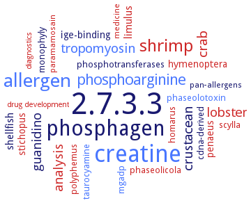
Word Map on EC 2.7.3.3

Reaction

ATP
+
L-arginine
=
ADP
+
Nomega-phospho-L-arginine

Synonyms

adenosine 5'-triphosphate-arginine phosphotransferase, adenosine 5'-triphosphate: L-arginine phosphotransferase, AK, AK-1, AK1, AK2, AK3, AK4, AK: L-arginine phosphotransferase, arginine kinase 1, arginine kinase 2, arginine kinase-1, arginine phosphokinase, ArgK, ARGK-2, ark, ATP: arginine N-phosphotransferase, ATP: arginine phosphotransferase, ATP: L-arginine phosphototransferase, ATP: L-arginine phosphotransferase, ATP:arginine N-phosphotransferase, ATP:arginine phosphotransferase, ATP:L-arginine N-phosphotransferase, ATP:L-arginine omega-N-phosphotransferase, ATP:L-arginine phosphotransferase, ESAK, F32B5.1, F44G3.2, F46H5.3, kinase, arginine (phosphorylating), McsB, MnAK2, MXAN2252 protein, PyAK1, PyAK2, PyAK3, PyAK4, rDer p 20, Tb09.160.4560, Tb927.9.6170, TbAK2, TbAK3, TcAK, TcAK1, TcAK2, W10C8.5, ZC434.8

ECTree

     2 Transferases
         2.7 Transferring phosphorus-containing groups
             2.7.3 Phosphotransferases with a nitrogenous group as acceptor
                2.7.3.3 arginine kinase

Crystallization

Crystallization on EC 2.7.3.3 - arginine kinase

Please wait a moment until all data is loaded. This message will disappear when all data is loaded.
CRYSTALLIZATION (Commentary)
ORGANISM
UNIPROT
LITERATURE
homology modeling and docking of inhibitors N-[2-(1H-imidazol-4-yl)ethyl]-3-[1-(2-methoxyethyl)indol-3-yl]propanamide, 5,7-dihydroxy-2-phenyl-6,8-bis(1-piperidylmethyl)chromen-4-one, (2S)-2-[[2-[[(2R)-2-[(2-phenoxyacetyl)amino]-3-phenylpropanoyl]amino]acetyl]amino]propanoic acid and 4-[[4-(1,3-benzodioxol-5-ylmethyl)piperazin-1-yl]methyl]-6,8-dimethyl-chromen-2-one. All inhibitors bind in the substrate binding pocket, all complexes maintain similar potential energy and are stable
hanging drop vapor diffusion method at 20°C. The crystal structure of H284A displays several structural changes, including the alteration of D324, a hydrogen-bonding network around H284, and the disruption of pi-stacking between the imidazole group of the H284 residue and the adenine ring of ATP
homology modeling and docking of inhibitors N-[2-(1H-imidazol-4-yl)ethyl]-3-[1-(2-methoxyethyl)indol-3-yl]propanamide, 5,7-dihydroxy-2-phenyl-6,8-bis(1-piperidylmethyl)chromen-4-one, (2S)-2-[[2-[[(2R)-2-[(2-phenoxyacetyl)amino]-3-phenylpropanoyl]amino]acetyl]amino]propanoic acid and 4-[[4-(1,3-benzodioxol-5-ylmethyl)piperazin-1-yl]methyl]-6,8-dimethyl-chromen-2-one. All inhibitors bind in the substrate binding pocket, all complexes maintain similar potential energy and are stable
-
crystal structures of mutant enzymes E314D at 1.9 A and E225Q at 2.8 A resolution shows that the precise alignment of substrates is subtly distorted
-
in complex with L-ornithine, L-citrulline, imino-L-ornithine, and D-arginine, hanging drop vapor diffusion method, using 26% (w/v) PEG 6000, 50 mM HEPES, and 100 mM MgCl2 at a pH of 8.0
polyethylene glycol precipitation of recombinant enzyme. Crystallization as a transition state analog
-
purified arginine kinase in binary complex with arginine and in a ternary transition state analogue complex, mixing of 20 mg/ml protein in 10 mM Tris-HCl, pH 8.0, and 1 mM DTT, with with 4 mM L-arginine, 0.001 ml of the solution is then mixed with 0.001 ml of crystallization solution containing 0.2 M sodium acetate, 0.1 M sodium cacodylate, pH 6.5, and 30% w/v PEG 8000, 16°C, 3 weeks. For the ternary complex, the enzyme is cocrystallized with ADP 16 mM, MgCl2 20 mM, L-arginine 40 mM, and NaNO3 75 mM, X-ray diffraction structure determination and analysis at 1.6-1.9 A resolution
hanging drop vapour diffusion method, the crystallographic structure of the alanine mutant at 2.3 A resolution, determined as a transition state analogue complex with arginine, nitrate, and MgADP2-, is nearly identical to wild type structure
Pleocyemata sp.
-
vapor-diffusion method, the apoenzyme and enzyme bound to arginine, both in the open conformation with the active site lid (residues 310-320) completely disordered. The guanidino group binding site in the apo structure appears to be organized to accept the arginine substrate
hanging drop vapor diffusion method at 20°C
homology modeling and docking of inhibitors N-[2-(1H-imidazol-4-yl)ethyl]-3-[1-(2-methoxyethyl)indol-3-yl]propanamide, 5,7-dihydroxy-2-phenyl-6,8-bis(1-piperidylmethyl)chromen-4-one, (2S)-2-[[2-[[(2R)-2-[(2-phenoxyacetyl)amino]-3-phenylpropanoyl]amino]acetyl]amino]propanoic acid and 4-[[4-(1,3-benzodioxol-5-ylmethyl)piperazin-1-yl]methyl]-6,8-dimethyl-chromen-2-one. All inhibitors bind in the substrate binding pocket, all complexes maintain similar potential energy and are stable
-
hanging drop method, structure of ligand-free TcAK is determined by molecular replacement methods and refined at 1.9 A resolution
hanging drop vapour diffusion method using 2.5 M ammonium sulfate, and 0.1 M Tris-HCl, pH 7.5