1.11.1.11: L-ascorbate peroxidase
This is an abbreviated version!
For detailed information about L-ascorbate peroxidase, go to the full flat file.

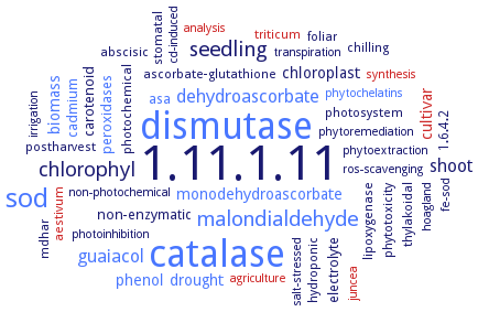
Word Map on EC 1.11.1.11 
-
1.11.1.11
-
catalase
-
dismutase
-
sod
-
malondialdehyde
-
seedling
-
chlorophyl
-
dehydroascorbate
-
guaiacol
-
shoot
-
drought
-
biomass
-
phenol
-
chloroplast
-
cultivar
-
monodehydroascorbate
-
electrolyte
-
carotenoid
-
non-enzymatic
-
cadmium
-
peroxidases
-
asa
-
lipoxygenase
-
thylakoidal
-
foliar
-
abscisic
-
photosystem
-
stomatal
-
hydroponic
-
postharvest
-
mdhar
-
aestivum
-
triticum
-
ascorbate-glutathione
-
photochemical
-
phytotoxicity
-
1.6.4.2
-
chilling
-
phytochelatins
-
irrigation
-
non-photochemical
-
cd-induced
-
phytoextraction
-
transpiration
-
salt-stressed
-
hoagland
-
photoinhibition
-
fe-sod
-
juncea
-
synthesis
-
analysis
-
agriculture
-
phytoremediation
-
ros-scavenging
- 1.11.1.11
- catalase
- dismutase
- sod
- malondialdehyde
- seedling
-
chlorophyl
- dehydroascorbate
- guaiacol
- shoot
- drought
- biomass
- phenol
- chloroplast
- cultivar
- monodehydroascorbate
-
electrolyte
-
carotenoid
-
non-enzymatic
- cadmium
- peroxidases
- asa
- lipoxygenase
- thylakoidal
-
foliar
-
abscisic
- photosystem
-
stomatal
-
hydroponic
-
postharvest
- mdhar
- aestivum
- triticum
-
ascorbate-glutathione
-
photochemical
-
phytotoxicity
-
1.6.4.2
-
chilling
- phytochelatins
-
irrigation
-
non-photochemical
-
cd-induced
-
phytoextraction
-
transpiration
-
salt-stressed
-
hoagland
-
photoinhibition
- fe-sod
- juncea
- synthesis
- analysis
- agriculture
-
phytoremediation
-
ros-scavenging
Reaction
2 L-ascorbate
+
Synonyms
Am-pAPX1, APEX2, APOX, APX, APX 1, APX 2, APX1, APX2, APX3, APX4, APX5, APX6, APX7, APx8, APXS, APXT, ascorbate peroxidase, ascorbate peroxidase 1, ascorbate peroxidase 2, ascorbate peroxidase 3, ascorbate peroxidase 4, ascorbate peroxidase 5, ascorbate peroxidase 6, ascorbate peroxidase 7, ascorbate peroxidase 8, ascorbate peroxidase6, ascorbic acid peroxidase, At1g07890, AT1G77490, AT3G09640, AT4G08390, AT4G32320, AT4G35000, AT4G35970, AtAPx08, AtAPX1, AtAPX2, AtAPX3, AtAPX5, AtAPX6, AtSAPX, AtstAPX, AtTAPX, cAPX, cAPX 2, CmstAPX, CrAPX4, CreAPX1, CreAPX2, CreAPX4, CreAPXheme, CsAPX1, cytoplasmic ascorbate peroxidase 1, cytosolic ascorbate peroxidase, GhAPX1, glyoxysomal APX, HvAPX1, L-ascorbate peroxidase, L-ascorbate peroxidase 3, L-ascorbate peroxidase 5, L-ascorbate peroxidase 6, L-ascorbate peroxidase, heme-containing, L-ascorbic acid peroxidase, L-ascorbic acid-specific peroxidase, LmAPX, MaAPX1, OsAPx1, OsAPx2, OsAPx3, OsAPx4, OsAPx5, OsAPx6, OsAPx7, OsAPx8, OsAPXa, OsAPXb, pAPX, Pavirv00022559m, peroxidase, ascorbate, peroxisomal ascorbate peroxidase, PgAPX1, PHYPA_001206, PHYPA_001884, PHYPA_021776, PHYPA_024580, PHYPA_024582, Potri.002G081900, Potri.004G174500, Potri.005G112200, Potri.005G161900, Potri.005G179200, Potri.006G089000, Potri.006G132200, Potri.006G254500, Potri.009G015400, Potri.009G134100, Potri.016G084800, PpAPX, PpAPX-S, PpAPX2, PpAPX2.1, PpAPX2.2, PpAPX3, PpAPX6-related, PtAPX-S.1, PtAPX-S.2, PtAPX-TL29, PtAPX.3, PtAPX1.1, PtAPX1.2, PtAPX2, PtAPX3, PtAPX5, PtAPX5-like, PtAPX6 related, PtotAPX, RcAPX, sAPX, stromal APX, stromal ascorbate peroxidase, stromal ascorbate peroxidases, TaAPX, tAPX, thylakoid ascorbate peroxidase, thylakoid membrane-bound ascorbate peroxidases, thylakoid-bound ascorbate peroxidase


 results (
 results (
 top
top


