1.3.1.6: fumarate reductase (NADH)
This is an abbreviated version!
For detailed information about fumarate reductase (NADH), go to the full flat file.
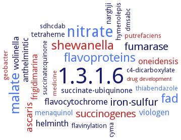
Word Map on EC 1.3.1.6
-
1.3.1.6
-
nitrate
-
malate
-
shewanella
-
fad
-
flavoproteins
-
fumarase
-
iron-sulfur
-
succinogenes
-
ascaris
-
flavocytochrome
-
helminth
-
oneidensis
-
wolinella
-
viologen
-
anthelmintic
-
frigidimarina
-
succinate-ubiquinone
-
tetraheme
-
menaquinol
-
narghji
-
thiabendazole
-
sdhcdab
-
c4-dicarboxylate
-
geobacter
-
flavinylation
-
succinate:quinone
-
dmsabc
-
hymenolepis
-
cyma
-
putrefaciens
-
medicine
-
drug development
- 1.3.1.6
- nitrate
- malate
- shewanella
- fad
- flavoproteins
- fumarase
-
iron-sulfur
- succinogenes
- ascaris
-
flavocytochrome
-
helminth
- oneidensis
-
wolinella
- viologen
-
anthelmintic
- frigidimarina
-
succinate-ubiquinone
-
tetraheme
- menaquinol
-
narghji
- thiabendazole
- sdhcdab
-
c4-dicarboxylate
- geobacter
-
flavinylation
-
succinate:quinone
- dmsabc
-
hymenolepis
-
cyma
- putrefaciens
- medicine
- drug development
Reaction
Synonyms
ABB37_00293, FRD, FRdABCD, FRDg, FRDm1, FRDm2, FRDS, Frds1p, fumarate reductase, KPA86010, KPK_2907, mitochondrial rhodoquinol-fumarate reductase, NADH-dependent fumarate reductase, NADH-FR, NADH-FRD, NADH-fumarate reductase, NFRD, QFR
ECTree

Advanced search results
Crystallization
Crystallization on EC 1.3.1.6 - fumarate reductase (NADH)
Please wait a moment until all data is loaded. This message will disappear when all data is loaded.
 top
topCRYSTALLIZATION (Commentary) 

ORGANISM
UNIPROT
LITERATURE
crystallized in the presence of octaethyleneglycol monododecyl ether and n-dodecyl-beta-D-maltopyranoside in a 3:2 weight ratio, crystals belongs to a orthorhombic space group with unit-cell parameters a=123.75 A, b= 29.08 A and c=221.12 A, diffracted to 2.8 A resolution using synchrotron radiation
-
mutant enzyme H505A and H505Y, hanging drop vapour diffusion method, protein solution: 6 mg/ml, 10 mM Tris-HCl, pH 8.5, well solution: 100 mM Tris-HCl, pH 7.4-8.2, 25°C, 80 mM NaCl, 16-19% PEG 8000, 10 mM fumarate, equal volume of 0.002 ml of protein solution and well solution, 10 days, cryoprotectant solution: 100 mM sodium acetate, pH 6.5, 20% PEG 8000, 10 mM fumarate, 80 mM NaCl, and 23% glycerol, X-ray diffraction structure determination and analysis at 1.8 A and 2.0 A resolution, respectively, molecular replacement
-
mutant enzyme H61A and H61M, hanging drop vapour diffusion method, protein solution: 6 mg/ml, 10 mM Tris-HCl, pH 8.5, well solution: 100 mM Tris-HCl, pH 7.4-8.5, 4°C, 80 mM NaCl, 16-19% PEG 8000, 10 mM fumarate, equal volume of 0.002 ml of protein solution and well solution, 10 days, cryoprotectant solution: 100 mM sodium acetate, pH 6.5, 20% PEG 8000, 10 mM fumarate, 80 mM NaCl, and 23% glycerol, X-ray diffraction structure determination and analysis at 2.1 A and 2.2 A resolution, respectively, molecular replacement
-