Any feedback?
Please rate this page
(all_enzymes.php)
(0/150)

BRENDA support

1.16.3.1: ferroxidase

This is an abbreviated version!
For detailed information about ferroxidase, go to the full flat file.
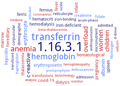
Word Map on EC 1.16.3.1

Reaction

4 Fe(II) + 4 H+ +

O2
= 4 Fe(III) + 2 H2O

Synonyms

AfFtn, apoferritin, bacterial ferritin, bacterial ferroxidase, bacterioferritin, bacterioferritin B, BFR, BfrB, blue copper oxidase, caeruloplasmin, ceruloplasmin, Cp115, Cp135, Cp200, CT1740, CtFtn, cyto-FOX, cytosolic FOX, DdBfr, Dpr, Dps, Dps protein, Dps-like peroxide resistance protein, Dps-Te, DpsA, DpsA-Te, DspA, EncA, encapsulin, encapsulin A, ferritin, ferro-O2-oxidoreductase, ferro:O2 oxidoreductase, ferroxidase, ferroxidase center of bacterioferritin, ferroxidase I, ferroxidase II, ferroxidase, iron II:oxygen oxidoreductase, Fet3, FET3 gene product, fet3p, FOX1, Ftn, FtnA, H ferritin, H' ferritin, H-chain ferritin, Helicobacter pylori neutrophil-activating protein, hephaestin, HP-NAP, HuHF, human ceruloplasmin form I, human H ferritin, human H-chain ferritin, iron(II): oxygen oxidoreductase, L-ferritin, M ferritin, MaDps, MCO1, MmcO, mnxDEFG, monophenol-o-monoxygenase, More, mouse ceruloplasmin, multicopper ferroxidase, multicopper oxidase, multicopper oxidase 1, multicopper oxidase CueO, multicopper oxidase-1, mushroom tyrosinase, mycobacterial multicopper oxidase, neutrophil-activating protein, non-ceruloplasmin ferroxidase, non-specific DNA-binding protein Dps/ferroxidase, rhHp, rHuHF, Rv0846c, serum ferroxidase, VcDps, VCE_000308, xanthine oxidoreductase

ECTree

     1 Oxidoreductases
         1.16 Oxidizing metal ions
             1.16.3 With oxygen as acceptor
                1.16.3.1 ferroxidase

Crystallization

Crystallization on EC 1.16.3.1 - ferroxidase

Please wait a moment until all data is loaded. This message will disappear when all data is loaded.
CRYSTALLIZATION (Commentary)
ORGANISM
UNIPROT
LITERATURE
structure of the ferritin at 2.1 and 2.7 A resolution for the native and iron bound proteins, respectively. X-ray crystallographic data indicate that the metal ion binding mode, secondary and tertiary structure of the enzyme closely resemble those of bacterial and eukaryotic H-type ferritins. However, the tetrahedral quarternary structure of the enzyme is unprecedented in its symmetry and in the presence of four large pores in the ferritin shell
hanging drop method, crystal structure of CueO at 1.1 A with the 45-residue methionine-rich segment fully resolved, revealing an N-terminal helical segment with methionine residues juxtaposed for Cu(I) ligation and a C-terminal highly mobile segment rich in methionine and histidine residues. Structures of CueO with a C500S mutation, and CueO with six methionines changed to serine
crystal structure analysis, PDB ID 1TJO
crystal structure determination of the enzyme apo form and metal-ion bound forms, such as iron, zinc, and cadmium, of HP-NAP, structure analysis
-
crystal structures of Zn2+- and Cd2+-bound forms of HP-NAP, and Cd2+-bound and apo forms of HP-NAP are determined: The coordination patterns of Zn2+ and Cd2+ are different but both metal ions can bind to the ferroxidase center (FOC), indicating that HP-NAP can store zinc and cadmium ions in addition to iron ions. Another zinc ion is found inside of the negatively-charged 3fold-related pore, as an iron ion in the iron-containing form, and therefore the pore is suitable for metal ions to pass through
purified recombinant H ferritin, 10 mg/ml protein in 10 mM Tris,HCl, pH 7.5, and 0.15 M NaCl, is mixed at equal volumes with crystallization solution containing 0.1 M BICINE-Na, pH 9.0, and 1.9-2.0 M MgCl2, 20°C, 1 week, X-ray diffraction structure determination and analysis at 1.52-1.97 A resolution
small angel X-ray scattering analysis of the complex with lactoferrin. Ceruloplasmin forms a 1:1 complex with lactoferrin. Complex formation occurs without major conformational rearrangements of either protein
in complex with zinc and terbium, at 1.8 A and 2.1 A resolution, respectively. Both ions bind to the ferroxidase center in the same location as iron
crystal structures of iron-loaded frog M ferritin determined by flash freezing crystals soaked for different times in iron(II) solutions under aerobic conditions. These structures provide the first X-ray picture of iron(III) products at the ferroxidase site in higher eukaryotes ferritins
purified recombinant iron-free E57A/E136A/D140A ferritin mutant variant, hanging drop vapor diffusion technique, mixing of 0.002 ml of 7 mg/ml protein in 20 mm Tris, pH 7.5, with 0.002 ml of reservoir solution composed of 1.6-2.0m MgCl2 and 0.1m bicine, pH 8.0, and equilibration against 0.6 ml reservoir solution, 8°C, 3–6 days to 10 days, for Fe2+-bound enzyme mutant, 0.1m bis-tris propane buffer at pH 6.5 is used for precipitation, X-ray diffraction structure determination and analysis at 1.50 A resolution, modeling
-
determined at 3 A. The crystallographic data implicate the importance of the extended C-terminal region in the iron entry from the three-fold channels to the ferroxidase centre and making iron more readily accessible for the oxidation
-
crystal structure analysis, PDB ID 4PT2
purified recombinant E57A/E136A/D140A mutant ferritin, X-ray diffraction structure determination and analysis
residues D283, E185, D409 provide a Fe(II) binding site that favors ferric ion thus reducing the reduction potential of the bound Fe(II). Residues E185 and D409 form part of the electron-transfer pathway from the bound Fe(II) to the protein’s type I Cu(II)
-
determination of the crystal structure of Streptococcus pyogenes Dpr in iron-free and iron-bound form at 2.0 and 1.93 A resolution, respectively
purified recombinant Dpr in complex with Zn2+, hanging drop vapour diffusion method, protein in 1 M succinic acid, 5% 2-propanol, and 1% w/v PEG 2000 MME, X-ray diffraction structure determination and analysis at 2.1 A resolution
-
purified recombinant getagged DpsA-Te, 0.001 ml of 10 mg/ml protein in 20 mM Tris-HCl, pH 7.5, is mixed with 0.001 ml of reservoir solution containing 12% w/v PEG 8000 in 0.1 m MES, pH 6.0, 2 weeks, X-ray diffraction structure determination and analysis at 2.4 A resolution, molecular replacement
Thermosynechococcus vestitus
crystal structure analysis, PDB ID 3DKT