2.7.1.107: diacylglycerol kinase (ATP)
This is an abbreviated version!
For detailed information about diacylglycerol kinase (ATP), go to the full flat file.
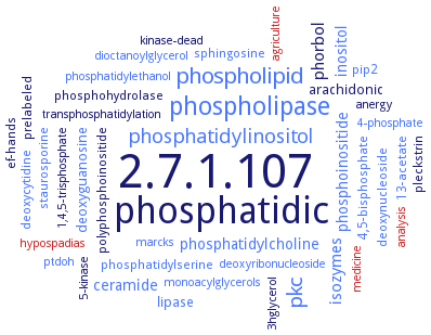
Word Map on EC 2.7.1.107
-
2.7.1.107
-
phosphatidic
-
phospholipase
-
phospholipid
-
pkc
-
phosphatidylinositol
-
phorbol
-
isozymes
-
phosphoinositide
-
phosphatidylcholine
-
inositol
-
ceramide
-
lipase
-
arachidonic
-
deoxyguanosine
-
deoxycytidine
-
ef-hands
-
phosphatidylserine
-
13-acetate
-
polyphosphoinositide
-
phosphohydrolase
-
prelabeled
-
deoxynucleoside
-
staurosporine
-
sphingosine
-
pip2
-
pleckstrin
-
4,5-bisphosphate
-
1,4,5-trisphosphate
-
4-phosphate
-
deoxyribonucleoside
-
analysis
-
marcks
-
kinase-dead
-
medicine
-
phosphatidylethanol
-
transphosphatidylation
-
dioctanoylglycerol
-
hypospadias
-
ptdoh
-
5-kinase
-
agriculture
-
3hglycerol
-
monoacylglycerols
-
anergy
- 2.7.1.107
-
phosphatidic
- phospholipase
- phospholipid
- pkc
- phosphatidylinositol
-
phorbol
- isozymes
- phosphoinositide
- phosphatidylcholine
- inositol
- ceramide
- lipase
-
arachidonic
- deoxyguanosine
- deoxycytidine
-
ef-hands
- phosphatidylserine
- 13-acetate
-
polyphosphoinositide
-
phosphohydrolase
-
prelabeled
- deoxynucleoside
- staurosporine
- sphingosine
- pip2
-
pleckstrin
- 4,5-bisphosphate
-
1,4,5-trisphosphate
- 4-phosphate
- deoxyribonucleoside
- analysis
- marcks
-
kinase-dead
- medicine
- phosphatidylethanol
-
transphosphatidylation
- dioctanoylglycerol
- hypospadias
- ptdoh
- 5-kinase
- agriculture
-
3hglycerol
- monoacylglycerols
-
anergy
Reaction
Synonyms
1,2-diacylglycerol kinase, 1,2-diacylglycerol kinase epsilon, adenosine 5'-triphosphate:1,2-diacylglycerol 3-phosphotransferase, arachidonoyl-specific diacylglycerol kinase, ATP:diacylglycerol phosphotransferase, DAG kinase, DAG-kinase, DAGK, DAGKalpha, DAGKepsilon, DG kinase, DGK, DGK zeta, DGK-1, DGK-3, DGK-3 diacylglycerol kinase, DGK-alpha, DGK-theta, DGK-zeta, DGK1, DGK2, DGK3, DGK4, DGK5, DGK6, DGK7, DGKA, DGKalpha, DgkB, DGKbeta, DGKdelta, DGKE, DGKepsilon, DGKeta, DGKeta1, DGKeta2, DGKeta3, DGKeta4, DGKgamma, DGKiota, DGKkappa, DGKksi, DGKtheta, DGKzeta, diacylglycerol kinase, diacylglycerol kinase (ATP dependent), diacylglycerol kinase 4, diacylglycerol kinase alpha, diacylglycerol kinase beta, diacylglycerol kinase delta, diacylglycerol kinase epsilon, diacylglycerol kinase eta, diacylglycerol kinase theta, diacylglycerol kinase zeta, diacylglycerol kinase-alpha, diacylglycerol kinase-epsilon, diacylglycerol kinase-zeta, diacylglycerol:ATP kinase, diglyceride kinase, epsilonDGK, eye-specific diacylglycerol kinase, kinase (phosphorylating), 1,2-diacylglycerol, kinase, 1,2-diacylglycerol (phosphorylating), OsDAGK1, Rv2252, sn-1,2-diacylglycerol kinase, type V DGK, YegS, YerQ
ECTree

Advanced search results
Engineering
Engineering on EC 2.7.1.107 - diacylglycerol kinase (ATP)
Please wait a moment until all data is loaded. This message will disappear when all data is loaded.
 top
topPROTEIN VARIANTS 

ORGANISM
UNIPROT
COMMENTARY 
LITERATURE
A722V
-
mutant isolated due to defects in DGK-1 controlled behaviour, altered behaviour compared to the wild-type enzyme, overview
C115Y
-
mutant isolated due to defects in DGK-1 controlled behaviour, altered behaviour compared to the wild-type enzyme, overview
C184Y
-
mutant isolated due to defects in DGK-1 controlled behaviour, altered behaviour compared to the wild-type enzyme, overview
G606E
-
mutant isolated due to defects in DGK-1 controlled behaviour, altered behaviour compared to the wild-type enzyme, overview
G609E
-
mutant isolated due to defects in DGK-1 controlled behaviour, altered behaviour compared to the wild-type enzyme, overview
G796R
-
mutant isolated due to defects in DGK-1 controlled behaviour, altered behaviour compared to the wild-type enzyme, overview
N745I
-
mutant isolated due to defects in DGK-1 controlled behaviour, altered behaviour compared to the wild-type enzyme, overview
P736S
-
mutant isolated due to defects in DGK-1 controlled behaviour, altered behaviour compared to the wild-type enzyme, overview
Q246stop
-
mutant isolated due to defects in DGK-1 controlled behaviour, altered behaviour compared to the wild-type enzyme, overview
Q422stop
-
mutant isolated due to defects in DGK-1 controlled behaviour, altered behaviour compared to the wild-type enzyme, overview
R167stop
-
mutant isolated due to defects in DGK-1 controlled behaviour, altered behaviour compared to the wild-type enzyme, overview
R180stop
-
mutant isolated due to defects in DGK-1 controlled behaviour, altered behaviour compared to the wild-type enzyme, overview
S880L
-
mutant isolated due to defects in DGK-1 controlled behaviour, altered behaviour compared to the wild-type enzyme, overview
W646stop
-
mutant isolated due to defects in DGK-1 controlled behaviour, altered behaviour compared to the wild-type enzyme, overview
W674stop
-
mutant isolated due to defects in DGK-1 controlled behaviour, altered behaviour compared to the wild-type enzyme, overview
W767stop
-
mutant isolated due to defects in DGK-1 controlled behaviour, altered behaviour compared to the wild-type enzyme, overview
G434D
-
kinase-defective dominant-negative mutant , impairs hepatocyte growth factor- and v-Src-induced cell scatter and migration, without affecting the loss of intercellular adhesions, impairs hepatocyre growth factor-induced cell spreading, lamellipodia formation, membrane ruffling, and focal adhesions remodeling and impairs hepatocyte growth factor-induced Rac activation and membrane targeting
Y335F
-
mutants is not tyrosine phosphorylated upon coexpression with activated Src mutant Y527F. Enzymatic activity is not stimulated by hepatocyte growth factor cell stimulation
A13K
site-directed mutagenesis, the mutant shows slightly reduced activity compared to the wild-type enzyme
A13R
site-directed mutagenesis, the mutant shows reduced activity compared to the wild-type enzyme
A14Q
-
significantly impaired catalytic function, without evidence of gross structural alterations, subunit mixing experiments of mutant enzymes, subunit mixing experiments of mutant enzymes
A30L
site-directed mutagenesis, the mutant shows 93% reduced activity compared to the wild-type enzyme
C46A/C113A
-
mutant lacking all Cys residues. Activity is slightly higher than wild-type
C46A/C113A/A29C
-
introduction of Cys residue at transmembrane helix 1 into mutant lacking the native Cys residues. Low activity mutant, 64% trimer formation compared to wild-type
C46A/C113A/A30C
-
introduction of Cys residue at transmembrane helix 1 into mutant lacking the native Cys residues. Low activity mutant, 79% trimer formation compared to wild-type
C46A/C113A/E28C
-
introduction of Cys residue at transmembrane helix 1 into mutant lacking the native Cys residues. Low activity mutant, 93% trimer formation compared to wild-type
C46A/C113A/Q33C
-
introduction of Cys residue at transmembrane helix 1 into mutant lacking the native Cys residues. Low activity mutant, 77% trimer formation compared to wild-type
C46A/C113A/R32C
-
introduction of Cys residue at transmembrane helix 1 into mutant lacking the native Cys residues. Low activity mutant, 63% trimer formation compared to wild-type
C46A/C113AE34C
-
introduction of Cys residue at transmembrane helix 1 into mutant lacking the native Cys residues. Low activity mutant, 100% trimer formation compared to wild-type
C46A/C113AF31C
-
introduction of Cys residue at transmembrane helix 1 into mutant lacking the native Cys residues. Low activity mutant, 72% trimer formation compared to wild-type
D80A
site-directed mutagenesis, the mutant shows highly reduced activity compared to the wild-type enzyme
D80E
site-directed mutagenesis, the mutant shows reduced activity compared to the wild-type enzyme
D80N
site-directed mutagenesis, the mutant shows reduced activity compared to the wild-type enzyme
D81A
site-directed mutagenesis, the mutant shows reduced activity compared to the wild-type enzyme
D81K
site-directed mutagenesis, the mutant shows reduced activity compared to the wild-type enzyme
D95A
site-directed mutagenesis, the mutant shows highly reduced activity compared to the wild-type enzyme
D95E
site-directed mutagenesis, the mutant shows reduced activity compared to the wild-type enzyme
D95N
E28A
site-directed mutagenesis, the mutation principally affects the binding of the Zn2+ ion, In the absence of the E28 side chain the zinc ions become purely coordinated by E76 and the ATP phosphates, the mutant shows highly reduced activity compared to the wild-type enzyme
E28D
site-directed mutagenesis, the mutant shows highly reduced activity compared to the wild-type enzyme
E28N
site-directed mutagenesis, the mutant shows highly reduced activity compared to the wild-type enzyme
E28Q
site-directed mutagenesis, the mutant shows highly reduced activity compared to the wild-type enzyme
E28R
site-directed mutagenesis, the mutant shows highly reduced activity compared to the wild-type enzyme
E34A
site-directed mutagenesis, the mutant shows reduced activity compared to the wild-type enzyme
E34D
site-directed mutagenesis, the mutant shows reduced activity compared to the wild-type enzyme
E34Q
site-directed mutagenesis, the mutant shows reduced activity compared to the wild-type enzyme
E69A
site-directed mutagenesis, inactive mutant, Asn72 plays a key role in catalysis. Its side-chain amide bridges Glu69 and Glu76
E69D
site-directed mutagenesis, inactive mutant, Asn72 plays a key role in catalysis. Its side-chain amide bridges Glu69 and Glu76
E69Q
site-directed mutagenesis, inactive mutant, Asn72 plays a key role in catalysis. Its side-chain amide bridges Glu69 and Glu76
E76A
E76D
site-directed mutagenesis, inactive mutant, Asn72 plays a key role in catalysis. Its side-chain amide bridges Glu69 and Glu76
E76L
-
significantly impaired catalytic function, without evidence of gross structural alterations, subunit mixing experiments of mutant enzymes
E76Q
site-directed mutagenesis, inactive mutant, Asn72 plays a key role in catalysis. Its side-chain amide bridges Glu69 and Glu76
G20A
site-directed mutagenesis, the mutant shows highly reduced activity compared to the wild-type enzyme
I110P
-
mutant enzyme can not be purified because its expression is toxic to the Escherichia coli host
I110R
-
mutant enzyme can not be purified because its expression is toxic to the Escherichia coli host
I110W
-
mutant is highly misfolding while at the same time being more stable than the wild-type protein
I110Y
-
mutant exhibits enhanced stability but folds with an efficiency similar to that of the wild type
K94A
K94 coordinates both alpha-phosphate and N7 of the adenine ring of ATP, the loss of the basic side-chain releases the adenine of ATP and the binding is lost, almost inactive mutant
K94L
-
significantly impaired catalytic function, without evidence of gross structural alterations. Km-value for MgATP2- raises 13fold, subunit mixing experiments of mutant enzymes
K94M
K94 coordinates both alpha-phosphate and N7 of the adenine ring of ATP, the loss of the basic side-chain releases the adenine of ATP and the binding is lost, the mutant shows highly reduced activity compared to the wild-type enzyme
K94R
K94 coordinates both alpha-phosphate and N7 of the adenine ring of ATP, the loss of the basic side-chain releases the adenine of ATP and the binding is lost, almost inactive mutant
N72A
site-directed mutagenesis, inactive mutant, Asn72 plays a key role in catalysis. Its side-chain amide bridges Glu69 and Glu76
N72D
site-directed mutagenesis, the mutant shows highly reduced activity compared to the wild-type enzyme
N72Q
site-directed mutagenesis, the mutant shows highly reduced activity compared to the wild-type enzyme
N72S
-
significantly impaired catalytic function, without evidence of gross structural alterations, subunit mixing experiments of mutant enzymes
R32A
site-directed mutagenesis, the mutant shows reduced activity compared to the wild-type enzyme
R32K
site-directed mutagenesis, the mutant shows reduced activity compared to the wild-type enzyme
R9A
site-directed mutagenesis, the mutant shows highly reduced activity compared to the wild-type enzyme
R9E
site-directed mutagenesis, the mutant shows highly reduced activity compared to the wild-type enzyme
R9H
site-directed mutagenesis, the mutant shows highly reduced activity compared to the wild-type enzyme
R9K
site-directed mutagenesis, the mutant shows highly reduced activity compared to the wild-type enzyme
S17A
site-directed mutagenesis, the mutant shows reduced activity compared to the wild-type enzyme
S73A
site-directed mutagenesis, the mutant shows reduced activity compared to the wild-type enzyme
S98A
site-directed mutagenesis, the mutant shows slightly reduced activity compared to the wild-type enzyme
W18L
-
site-directed mutagenesis, reduced activity compared to the wild-type enzyme
W18L/W25L
-
site-directed mutagenesis, reduced activity compared to the wild-type enzyme
W18L/W25L/W47L
-
site-directed mutagenesis, reduced activity compared to the wild-type enzyme
W18L/W25L/W47L/W117L
-
site-directed mutagenesis, reduced activity compared to the wild-type enzyme
W18L/W47L/W117L
-
site-directed mutagenesis, reduced activity compared to the wild-type enzyme
W25L
-
site-directed mutagenesis, reduced activity compared to the wild-type enzyme
Y86A
site-directed mutagenesis, the mutant shows unaltered activity compared to the wild-type enzyme
Y86F
site-directed mutagenesis, the mutant shows slightly reduced activity compared to the wild-type enzyme
E35G
-
mutant exhibits greatly reduced polymerization. Samples of the mutant incubated with an excess of zinc are shifted entirely to the insoluble fraction. In absence of zinc, most of the mutant protein sample is monomeric. In the presence of added zinc, the mutant organizes into large sheet structures
F369A/F372A
-
significant decrease in diacylglycerol kinase activity. Mutant cells display reduced uptake of transferrin
F369A/F372A/F748A
-
significant decrease in diacylglycerol kinase activity. Mutant cells display reduced uptake of transferrin
F748A
-
diacylglycerol kinase activity similar to that of wild-type. Mutant cells display reduced uptake of transferrin
G236R
-
site-directed mutagenesis, highly reduced activity compared to the wild-type isozyme theta
H3A/C20A/H38A/C60A
-
mutant shows a reduced zinc retention of 3%. Construct does not show any increase in turbidity after incubation with 50 microM zinc acetate. In the absence of zinc, short polymers are observed, much like the wild-type protein. When zinc is added, polymers increase in prevalence and length marginally but no large sheet structures are formed in 50 microM zinc. Mutant diminishes the formation of cytoplasmic puncta, shows partially impaired regulation of transport to the plasma membrane, and lacks the ability to inhibit the formation of CopII coated vesicles
L241V
-
site-directed mutagenesis, slightly reduced activity compared to the wild-type isozyme theta
L447
residue is required for the cholesterol recognition/interaction amino acid consensus motif, mutation results in a loss of enzymatic activity
L447I
ratio of enzymic activity with substrates 1-stearoyl-2-linoleoyl-sn-glycerol to 1-stearoyl-2-arachidonoyl-sn-glycerol is 0.054
P244A
-
site-directed mutagenesis, reduced activity compared to the wild-type isozyme theta
P244L
-
site-directed mutagenesis, reduced activity compared to the wild-type isozyme theta
P245L
-
site-directed mutagenesis, highly reduced activity compared to the wild-type isozyme theta
P32A
redcution of both Km and kcat value, while maintianing the ratio kcat/Km constant. Specificity of mutant for substrates with polyunsaturated acyl chains is retained. Mutant has a higher affinity for membranes
R457K
ratio of enzymic activity with substrates 1-stearoyl-2-linoleoyl-sn-glycerol to 1-stearoyl-2-arachidonoyl-sn-glycerol is 0.217
R457Q
mutation results in the loss of the cholesterol recognition/interaction amino acid consensus motif and the loss of a positively charged residue, resulting in a higher enzymatic activity than wild-type. Ratio of enzymic activity with substrates 1-stearoyl-2-linoleoyl-sn-glycerol to 1-stearoyl-2-arachidonoyl-sn-glycerol is 0.099. Mutant gains preference for substrate 1-stearoyl-2-docosahexaenoyl-sn-glycerol
S240T
-
site-directed mutagenesis, activity is unaltered compared to the wild-type isozyme theta
S258D/S265D/S270D/S271D
-
mutation in diacylglycerol kinase zeta for mimicking of protein kinase C phosphorylation of serine residues within the MARCKS phosphorylation site domain. Mutations do prevent binding to retinoblastoma protein
S258N/S265N/S270N/S271N
-
mutation in diacylglycerol kinase zeta for mimicking of protein kinase C phosphorylation of serine residues within the MARCKS phosphorylation site domain. Mutations do not prevent binding to retinoblastoma protein and subsequent stimulation of activity
V52E
-
mutant exhibits greatly reduced polymerization, no polymers are visible in zinc-free conditions. After zinc addition, large sheet structures appear
Y335F
expression of wild-type diacylglycerol kinase alpha markedly reduces ERK phosphorylation, whereas the effect of expressing the nonphosphorylatableY335F mutant is much less pronounced
Y451F
mutation results in a loss of a hydroxyl group and an essential residue of the cholesterol recognition/interaction amino acid consensus motif, leading to a higher activity than the wild-type protein. Ratio of enzymic activity with substrates 1-stearoyl-2-linoleoyl-sn-glycerol to 1-stearoyl-2-arachidonoyl-sn-glycerol is 0.107. Mutant gains preference for substrate 1,2-diarachidonoyl-sn-glycerol, with activities comparable to 1-stearoyl-2-arachidonoyl-sn-glycerol
T57P
-
mutant with reduced activity, used for construcution of fusion protein for genetic selection of soluble mutants
G279D
inactive mutant of diacylglycerol kinase epsilon due to replacement of ATP-binding domain GxGxxG with GxDxxG. Similar subcellular localization as wild type
G356D
inactive mutant of diacylglycerol kinase zeta due to replacement of ATP-binding domain GxGxxG with GxDxxG. Similar subcellular localization as wild type
G428D
inactive mutant of diacylglycerol kinase alpha due to replacement of ATP-binding domain GxGxxG with GxDxxG. Similar subcellular localization as wild type
G491D
inactive mutant of diacylglycerol kinase gamma due to replacement of ATP-binding domain GxGxxG with GxDxxG. Similar subcellular localization as wild type
G495D
inactive mutant of diacylglycerol kinase beta due to replacement of ATP-binding domain GxGxxG with GxDxxG. In contrast to the filamentous image of the wild type, mutant is diffusely distributed throughout the cytoplasm
D124A
D271A
D68A
E273A
D497N
-
site-directed mutagenesis, 0.9% of wild-type activity, reduced stimulation by Ca2+ and phosphatidylserine compared to the wild-type enzyme
D529N
-
site-directed mutagenesis, 5.5% of wild-type activity, unaltered stimulation by Ca2+ and phosphatidylserine compared to the wild-type enzyme
D697N
-
site-directed mutagenesis, 4.0% of wild-type activity, reduced stimulation by Ca2+ and phosphatidylserine compared to the wild-type enzyme
additional information
D95N

-
significantly impaired catalytic function, without evidence of gross structural alterations. Km-value for MgATP2- raises 18fold, subunit mixing experiments of mutant enzymes
D95N
site-directed mutagenesis, the mutant shows unaltered activity compared to the wild-type enzyme
E76A
site-directed mutagenesis, inactive mutant, Asn72 plays a key role in catalysis. Its side-chain amide bridges Glu69 and Glu76
E76A
site-directed mutagenesis, the mutation principally affects the binding of the Zn2+ ion
D124A
residue involved in the Mg1-water network, no catalytic acitivity
D271A
residue involved in the Mg1-water network, no catalytic acitivity
D68A
no catalytic activity. Role of D68 in mediating the interaction of Mg2+ with the gamma-phosphate of ATP
additional information
-
expression of fusion constructs of Green Fluorescent Protein to truncated forms of diacylglycerol kinase 1 and diacylglycerol kinase 2 missing the catalytic and accessory domains. Fusion proteins are localized to the endoplasmic reticulum. Fusion constructs of N-terminal 50 amino acid residues of diacylglycerol kinase 1 and the 43 residues of diacylglycerol kinase 2 to the Yellow Fluorescent Protein alos localize to the endoplasmic reticulum
additional information
-
functional complementation of Escherichia coli dgkA mutant. Conditional inactivation of gene expression leads to the accumulation of diacylglycerol and the cessation of lipoteichoic acid formation in Bacillus subtilis
additional information
functional complementation of Escherichia coli dgkA mutant. Conditional inactivation of gene expression leads to the accumulation of diacylglycerol and the cessation of lipoteichoic acid formation in Bacillus subtilis
additional information
-
determination of mutational defects/molecular lesions affecting the enzyme activity and splice forms of the enzyme, overview
additional information
-
deletion mutants lacking respectively the entire C-terminal half of diacylglycerol kinase alpha or the last 13 amino acids PPPRSTNFFGFLS. Contrary to wild-type, mutants are not pulled down by immobilized GST-Src-SH3 fusion protein
additional information
-
downregulation of diacylglycerol kinase alpha by siRNA impairs hepatocyte growth factor- and v-Src-induced cell scatter and migration, without affecting the loss of intercellular adhesions, impairs hepatocyre growth factor-induced cell spreading, lamellipodia formation, membrane ruffling, and focal adhesions remodeling and impairs hepatocyte growth factor-induced Rac activation and membrane targeting
additional information
-
the ATP binding sequence of isozyme DGK2in strain rdgA contains the mutant GXDXXG motif leading to rapid retinal degeneration after birth
additional information
generation of a mutant defective in the gene encoding DGK, rdgA
additional information
-
generation of a mutant defective in the gene encoding DGK, rdgA
additional information
-
generation of a mutant defective in the gene encoding DGK, rdgA
-
additional information
construction of a thermostabilized DELTA4 (4 changes relative to wild-type) form of DgkA using the DELTA7 structure, overview
additional information
-
construction of deletion mutants, N- or C-terminal truncations inactivate the isozyme theta
additional information
-
mutation of the second glycine in the binding sequence motif GXGXXG of the ATP-binding site to aspartate or alanine renders the mutant enzymes catalytically inactive
additional information
-
truncated form of the protein (DGKDELTAepsilon) lacking the 40 N-terminal amino acids. Full-length FLAG-DGKepsilon and truncated FLAG-DGKepsilon are both more specific for 1-stearoyl-2-arachidonoyl-sn-glycerol than for 1,2-dioleoyl-sn-glycerol. 1-Stearoyl-2-linoleoyl-sn-glycerol exhibits intermediate specificity for both forms of the enzyme. The truncated form of the enzyme maintains substrate specificity for lipids with an arachidonoyl moiety present at the sn-2 position. The truncation increases the catalytic rate constant for all three substrates and may suggest a role in the negative regulation of this enzyme
additional information
-
diacylglycerol kinase delta has alternative splicing variants, type 1 DGKdelta1 and type 2 DGKdelta2, with calculated molecular masses of 130 and 134 kDa, respectively. HeLa cells express both type 1 and 2 DGKdelta, and COS7 cells express only type 2 DGKdelta. In DGKdelta-knockdown cells uptake of transferrin is reduced. DGKdelta2 is partially co-localized with clathrin or adaptor protein AP2alpha in COS7 cells. Mutants lacking binding ability to AP2alpha as well as kinase-negative mutants cannot compensate for the uptake of transferrin inhibited by siRNA treatment. Overexpression of wild-type diacylglycerol kinase delta2 completely recovers the transferrin uptake
additional information
expression of a peptide corresponding to a putative transmembrane segment which comprises approximately residues 20-40 and is found in all forms of mammalian diacylglycerol kinase epsilon. Peptide KKKKLILWTLCSVLLPVFITFWKKKKK-NH2 has increased helical content and significant blue shifts in the presence of anionic but not zwitterionic bilayer membranes. Peptide dimerizes and preferentially interacts with cholesterol in lipid films comprised of homogeneous mixtures of cholesterol and phosphatidylcholine, yet the presence of cholesterol in hydrated vesicle bilayers decreases its helical content
additional information
-
expression of truncated FLAG-tagged protein lacking the 40 N-terminal amino acids, which includes the hydrophobic segment. Truncated protein maintains substrate specificity and increases catalytic rate constant. Truncated protein may be extracted with 1.5 M KCl at neutral pH value, while wild-type protein remains fully membrane bound
additional information
heterologous expression of diacylglycerol kinase gamma in COS-7 cells. Upon stimulation with epidermal growth factor, enzyme specifically interacts and co-localizes at the plasma membrane with beta2-chimaerin. Enzyme enhances epidermal growth factor-dependent translocation of beta2-chimaerin to the plasma membrane and markedly augments epidermal growth factor-dependent GTPase-activating protein activity of beta2-chimaerin
additional information
in a female patient with a de novo balanced translocation, 46,X,t(X,2)(p11.2,q37)dn, who exhibits seizures, capillary abnormality, developmental delay, infantile hypotonia, and obesity, diacylglycerol kinase delta is disrupted at 2q37. Diacylglycerol kinase delta is involved in the etiology of seizures
additional information
-
overexpression of wild-type diacylglycerol kinase alpha, but not of its kinase-dead mutant, markedly suppresses tumor necrosis factor alpha-induced apoptosis of AKI human melanoma cells and enhances the tumor necrosis factor alpha-stimulated transcriptional activity of transcription factor NF-kappaB. siRNA-mediated knock-down of diacylglycerol kinase alpha enhances the apoptosis. Overexpression of isoforms beta and gamma has no detectable effect on apoptosis
additional information
-
RNA interference-mediated knockdown of diacylglycerol kinase zeta leads to accelerated transferrin receptor exit from the lymphocyte endocytic recycling compartment back to the plasma membrane
additional information
RNA interference-mediated knockdown of diacylglycerol kinase zeta leads to accelerated transferrin receptor exit from the lymphocyte endocytic recycling compartment back to the plasma membrane
additional information
-
silencing of diacylglycerol kinase theta by siRNA expression inhibits cAMP-dependent CYP17 transcription. LXXLL motifs in diacylglycerol kinase theta mediate a direct interaction of steroidogenic factor 1 with the kinase and may facilitate binding of phosphatidic acid to the receptor
additional information
-
mutation of the second glycine in the binding sequence motif GXGXXG of the ATP-binding site to aspartate or alanine renders the mutant enzymes catalytically inactive
additional information
construction of mutant protein lacking the regulatory N-terminus and both C1 domains. Mutant is enzymatically active. deletion analysis reveals that the C1 domains are essential for plasma membrane targeting in intact cells but unnecessary for catalytic activity. The C-terminal sequence is required for membrane-binding in a phosphatidic acid-dependent manner. In the absence of the calcium binding domain, receptor-dependent translocation of the truncated protein is regulated by phosphorylation of residue Y335
additional information
-
creation of thoracic transverse aortic constriction in transgenic mice with cardiac-specific overexpression of diacylglycerol kinase zeta and wild-type mice. Increases in heart weight at 4 weeks after thoracic transverse aortic constriction are attenuated in diacylglycerol kinase zeta transgenic mice compared with wild-type mice. Increases in interventricular septal thickness, dilatation of the left ventricular cavity, and decreases in left ventricular systolic function in wild-type mice are observed at 4 weeks after surgery and are attenuated in diacylglycerol kinase zetatransgenic mice. Contrary to wild-type, cardiac fibrosis and gene induction of type I and type III collagens, but not transforming growth factor are blocked in diacylglycerol kinase zeta transgenic mice
additional information
-
deficiency for diacylglycerol kinase zeta results in impaired interleukin 12 and tumor necrosis factor alpha production following toll-like receptor stimulation in vitro and in vivo, increased resistance to endotoxin shock, and enhanced susceptibility to Toxoplasma gondii infection. Deficiency results in increased activation of the phosphatidylinositol 3-kinase-Akt pathway. Inhibition of phosphatidylinositol 3-kinase activity or treatment with phosphatidic acid can restore lipopolysaccharide-induced interleukin 12 production by diacylglycerol kinase zeta-deficient mice
additional information
-
diacylglycerol kinase delta knock-down mice reveal abnormal epiletic discharges and electrographic seizures in three out of six homozygotes
additional information
-
expression of isolated His-tagged sterile alpha-motif domain of diacylglycerol kinase delta1, comprised of residues 1097-1164. The domain forms large aggregates with a molecular weight of 250000 Dalton or greater, while calculated monomer molecular weight is 9500 Dalton
additional information
generation of double transgenic mice with cardiac-specific overexpression of both diacylglycerol kinase zeta and G-protein subunit alphaq. Diacylglycerol kinase zeta prevents cardiac dysfunction, determined by dilatation of left ventricular dimensions, reduction of left ventricular fractional shortening, and marked increases in left ventricular end-diastolic pressure in G-protein subunit alphaq transgenic mice. Translocation of protein kinase C isoforms, phosphorylation activity of c-jun N-terminal kinase and p38 mitogen-activated protein kinase in G-protein subunit alphaq transgenic mice are attenuated by diacylglycerol kinase zeta?. Diacylglycerol kinase zeta improves the survival rate of G-protein subunit alphaq transgenic mice
additional information
-
generation of heterozygous diacylglycerol kinase delta whole-body knockout mice. Diacylglycerol kinase delta haploinsufficiency increases diacylglycerol content, reduces peripheral insulin sensitivity, insulin signaling, and glucose transport, and leads to age-dependent obesity. Metabolic flexibility is impaired
additional information
-
generation of mice lacking both diacylglycerol kinase alpha and zeat. Absence of both diacylglycerol kinases results in a severe decrease in the number of CD4+CD8- and CD4-CD8+ single-positive thymocytes correlating with increased diacylglycerol kinase-mediated signaling. Positive selection, but not negative selection, is impaired in diacylglycerol kinase alpha-/- zeta -/- mice. The developmental blockage in diacylglycerol kinase alpha-/- zeta -/- mice can be partially overcome by treatment with phosphatidic acid. Decreased diacylglycerol kinase activity also promotes thymic lymphomagenesis accompanying elevated Ras and Erk1/2 activation
additional information
in mouse ventricular myocytes overexpressing diaclyglycerol kinase zeta, the effect induced by endothelin-1 on Ca2+ transients and cell shortening are abolished
additional information
-
in mouse ventricular myocytes overexpressing diaclyglycerol kinase zeta, the effect induced by endothelin-1 on Ca2+ transients and cell shortening are abolished
additional information
-
overexpression of diacylglycerol kinase zeta blocks cells in G1 phase of cell cycle. Cell cycle arrest is accompanied by decreased levels of retinoblastoma protein phosphorylated on Ser-807/811. Down-regulation by siRNA increases the number of cells in both the S and G2/M phases of the cell cycle and prevents the cell cycle block characterizing C2C12 cell myogenic differentiation
additional information
transgenic mice with cardiac-specific overexpression of diacylglycerol kinase zeta. Left ventricular chamber dilatation, reduction of left ventricular systolic function and increases in left ventricular weight and lung weight at 4 weeks after myocardial infarction are attenuated in transgenic mice compared with wild-type mice. In the noninfarct area, fibrosis fraction and upregulation of profibrotic genes, such as transforming growth factor-1, collagen type I, and collagen type III, are blocked in transgenic mice. Survival rate at 4 weeks after myocardial infarction is higher in transgenic mice than in wild-type
additional information
-
transgenic mice with cardiac-specific overexpression of diacylglycerol kinase zeta. Left ventricular chamber dilatation, reduction of left ventricular systolic function and increases in left ventricular weight and lung weight at 4 weeks after myocardial infarction are attenuated in transgenic mice compared with wild-type mice. In the noninfarct area, fibrosis fraction and upregulation of profibrotic genes, such as transforming growth factor-1, collagen type I, and collagen type III, are blocked in transgenic mice. Survival rate at 4 weeks after myocardial infarction is higher in transgenic mice than in wild-type
additional information
nuclear export signal mutant forms of DGK-zeta accumulate in the nucleus to a much greater extent than wild type DGK-zeta
additional information
-
nuclear export signal mutant forms of DGK-zeta accumulate in the nucleus to a much greater extent than wild type DGK-zeta
additional information
-
generation of enzyme-deficient DGKdelta-KO mouse embryonic fibroblasts
additional information
generation of transgenic tobacco plants constitutively expressing rice diacylglycerol kinase. Overexpression results in enhanced resistance against infection by tobacco mosaic virus and Phytophthora parasitica var. nicotianae
additional information
-
generation of transgenic tobacco plants constitutively expressing rice diacylglycerol kinase. Overexpression results in enhanced resistance against infection by tobacco mosaic virus and Phytophthora parasitica var. nicotianae
additional information
-
antisense silencing of diacylglacerol kinase isoform delta, but not alpha, expression is sufficient to prevent the effect of high glucose on protein kinase alpha activity, insulin receptor signaling, and glucose uptake
additional information
-
diacylglycerol kinase zeta siRNA tranfection decreases H2O2-induced apoptosis. Overexpression of kinase-dead diacylglycerol kinase zeta significantly increases protein kinase D phosphorylation
additional information
-
construction of deletion mutants DELTA196, lacking the RVH motif and the EF hand, and DELTA332, lacking the RVH motif, the EF hand, and the C1 domain, mutant DELTA332 shows 50% of wild-type activity