1.1.1.3: homoserine dehydrogenase
This is an abbreviated version!
For detailed information about homoserine dehydrogenase, go to the full flat file.
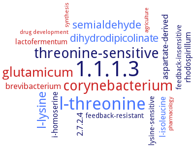
Word Map on EC 1.1.1.3
-
1.1.1.3
-
l-threonine
-
threonine-sensitive
-
corynebacterium
-
glutamicum
-
l-lysine
-
semialdehyde
-
dihydrodipicolinate
-
l-isoleucine
-
brevibacterium
-
aspartate-derived
-
2.7.2.4
-
i-homoserine
-
rhodospirillum
-
lysine-sensitive
-
lactofermentum
-
feedback-insensitive
-
feedback-resistant
-
synthesis
-
drug development
-
agriculture
-
pharmacology
- 1.1.1.3
- l-threonine
-
threonine-sensitive
- corynebacterium
- glutamicum
- l-lysine
- semialdehyde
- dihydrodipicolinate
- l-isoleucine
- brevibacterium
-
aspartate-derived
-
2.7.2.4
-
i-homoserine
-
rhodospirillum
-
lysine-sensitive
- lactofermentum
-
feedback-insensitive
-
feedback-resistant
- synthesis
- drug development
- agriculture
- pharmacology
Reaction
Synonyms
AK-HDH, AK-HSD-1, AK-HSDH, AK-HseDH, aspartate kinase-homoserine dehydrogenase, aspartokinase-homoserine dehydrogenase I, bifunctional aspartate kinase-homoserine dehydrogenase, BsHSD, HDH, hom, hom-1, Hom6, homoserine dehydrogenase 1, homoserine dehydrogenase 2, HSD, HSDH, HseDH, More, orf19.2951, PbHSD, SACOL1362, StHSD, thrA, TM_0547, TTHA0489, TtHSD
ECTree

Advanced search results
General Information
General Information on EC 1.1.1.3 - homoserine dehydrogenase
Please wait a moment until all data is loaded. This message will disappear when all data is loaded.
 top
topGENERAL INFORMATION 

ORGANISM
UNIPROT
COMMENTARY 
LITERATURE
evolution
malfunction
metabolism
physiological function
additional information
evolution

the catalytic region of the enzyme is unique, the nucleotide-binding domain conforms to the Rossmann fold-like conventional NAD(P)-dependent dehydrogenases
evolution
enzyme homoserine dehydrogenase (HSD) belongs to the family of oxidoreductases and is essential for the biosynthesis of threonine (Thr), methionine (Met), and lysine (Lys) in the metabolic pathway of fungi and plants
evolution
enzyme homoserine dehydrogenase (HSD) belongs to the family of oxidoreductases and is essential for the biosynthesis of threonine (Thr), methionine (Met), and lysine (Lys) in the metabolic pathway of fungi and plants
evolution
the orientation of the three domains in the bifunctional aspartate kinase-homoserine dehydrogenase (AK-HseDH) homologue found in Thermotoga maritima totally differs from those observed in previously known AK-HseDHs, the domains line up in the order HseDH, AK, and regulatory domain
evolution
-
enzyme homoserine dehydrogenase (HSD) belongs to the family of oxidoreductases and is essential for the biosynthesis of threonine (Thr), methionine (Met), and lysine (Lys) in the metabolic pathway of fungi and plants
-
evolution
-
the orientation of the three domains in the bifunctional aspartate kinase-homoserine dehydrogenase (AK-HseDH) homologue found in Thermotoga maritima totally differs from those observed in previously known AK-HseDHs, the domains line up in the order HseDH, AK, and regulatory domain
-
evolution
-
enzyme homoserine dehydrogenase (HSD) belongs to the family of oxidoreductases and is essential for the biosynthesis of threonine (Thr), methionine (Met), and lysine (Lys) in the metabolic pathway of fungi and plants
-
evolution
-
the catalytic region of the enzyme is unique, the nucleotide-binding domain conforms to the Rossmann fold-like conventional NAD(P)-dependent dehydrogenases
-
evolution
-
the orientation of the three domains in the bifunctional aspartate kinase-homoserine dehydrogenase (AK-HseDH) homologue found in Thermotoga maritima totally differs from those observed in previously known AK-HseDHs, the domains line up in the order HseDH, AK, and regulatory domain
-
evolution
-
the orientation of the three domains in the bifunctional aspartate kinase-homoserine dehydrogenase (AK-HseDH) homologue found in Thermotoga maritima totally differs from those observed in previously known AK-HseDHs, the domains line up in the order HseDH, AK, and regulatory domain
-
malfunction
HOM6 deletions cause translational arrest in cells grown under amino acid starvation conditions. HOM6 deletion reduces Candida albicans cell adhesion to polystyrene, which is a common plastic used in many medical devices. HOM6-homozygous mutants are hypersensitive to hygromycin B and cycloheximide as compared with wild-type, HOM6-heterozygous, and HOM6-reintegrated strains. HOM6 deletion affects translation and leads to the accumulation of free ribosomes
malfunction
the chemical mutagen ENU causes a mutation in the homoserine serine dehydrogenase enzyme which diverted the aspartyl-beta-semialdehyde to bind with 2,3-dihydrodipicolinate synthase to participate in the L-lysine synthesis through 2,3 meso-diaminopimelate (Meso-Dap). Being a recombinant for diaminopimelate dehydrogenase (ddh), the auxotrophic mutant for the homoserine dehydrogenase follows the ddh pathway by overexpression of ddh by deviating the acetyltransferase and succinyl transferase is the reason for the high yield of L-lysine production
malfunction
-
the chemical mutagen ENU causes a mutation in the homoserine serine dehydrogenase enzyme which diverted the aspartyl-beta-semialdehyde to bind with 2,3-dihydrodipicolinate synthase to participate in the L-lysine synthesis through 2,3 meso-diaminopimelate (Meso-Dap). Being a recombinant for diaminopimelate dehydrogenase (ddh), the auxotrophic mutant for the homoserine dehydrogenase follows the ddh pathway by overexpression of ddh by deviating the acetyltransferase and succinyl transferase is the reason for the high yield of L-lysine production
-
malfunction
Candida albicans SC5314 / ATCC MYA-2876
-
HOM6 deletions cause translational arrest in cells grown under amino acid starvation conditions. HOM6 deletion reduces Candida albicans cell adhesion to polystyrene, which is a common plastic used in many medical devices. HOM6-homozygous mutants are hypersensitive to hygromycin B and cycloheximide as compared with wild-type, HOM6-heterozygous, and HOM6-reintegrated strains. HOM6 deletion affects translation and leads to the accumulation of free ribosomes
-
malfunction
-
the chemical mutagen ENU causes a mutation in the homoserine serine dehydrogenase enzyme which diverted the aspartyl-beta-semialdehyde to bind with 2,3-dihydrodipicolinate synthase to participate in the L-lysine synthesis through 2,3 meso-diaminopimelate (Meso-Dap). Being a recombinant for diaminopimelate dehydrogenase (ddh), the auxotrophic mutant for the homoserine dehydrogenase follows the ddh pathway by overexpression of ddh by deviating the acetyltransferase and succinyl transferase is the reason for the high yield of L-lysine production
-
malfunction
-
the chemical mutagen ENU causes a mutation in the homoserine serine dehydrogenase enzyme which diverted the aspartyl-beta-semialdehyde to bind with 2,3-dihydrodipicolinate synthase to participate in the L-lysine synthesis through 2,3 meso-diaminopimelate (Meso-Dap). Being a recombinant for diaminopimelate dehydrogenase (ddh), the auxotrophic mutant for the homoserine dehydrogenase follows the ddh pathway by overexpression of ddh by deviating the acetyltransferase and succinyl transferase is the reason for the high yield of L-lysine production
-
metabolism
homoserine dehydrogenase (HSD) is an oxidoreductase in the aspartic acid pathway. The L-homoserine produced by this enzyme at the first branch point of the aspartic acid pathway is a precursor for essential amino acids such as L-threonine, L-methionine and L-isoleucine
metabolism
homoserine dehydrogenase (HSD) is an oxidoreductase that is involved in the reversible conversion of L-aspartate semialdehyde to L-homoserine in a dinucleotide cofactor-dependent reduction reaction. HSD is thus a crucial intermediate enzyme linked to the biosynthesis of several essential amino acids such as lysine, methionine, isoleucine and threonine
metabolism
homoserine dehydrogenase activity and is involved in the biosynthesis of methionine and threonine
metabolism
homoserine dehydrogenase is a key enzyme in the L-threonine pathway
metabolism
biosynthetic pathway from L-aspartate to L-homoserine involving the bifunctional enzyme, overview
metabolism
homoserine dehydrogenase (HSD) catalyzes the reversible conversion of L-aspartate-4-semialdehyde to L-homoserine in the aspartate pathway for the biosynthesis of lysine, methionine, threonine, and isoleucine
metabolism
-
homoserine dehydrogenase (HSD) catalyzes the reversible conversion of L-aspartate-4-semialdehyde to L-homoserine in the aspartate pathway for the biosynthesis of lysine, methionine, threonine, and isoleucine
-
metabolism
-
biosynthetic pathway from L-aspartate to L-homoserine involving the bifunctional enzyme, overview
-
metabolism
-
homoserine dehydrogenase (HSD) is an oxidoreductase in the aspartic acid pathway. The L-homoserine produced by this enzyme at the first branch point of the aspartic acid pathway is a precursor for essential amino acids such as L-threonine, L-methionine and L-isoleucine
-
metabolism
-
homoserine dehydrogenase (HSD) is an oxidoreductase that is involved in the reversible conversion of L-aspartate semialdehyde to L-homoserine in a dinucleotide cofactor-dependent reduction reaction. HSD is thus a crucial intermediate enzyme linked to the biosynthesis of several essential amino acids such as lysine, methionine, isoleucine and threonine
-
metabolism
-
homoserine dehydrogenase is a key enzyme in the L-threonine pathway
-
metabolism
Candida albicans SC5314 / ATCC MYA-2876
-
homoserine dehydrogenase activity and is involved in the biosynthesis of methionine and threonine
-
metabolism
-
biosynthetic pathway from L-aspartate to L-homoserine involving the bifunctional enzyme, overview
-
metabolism
-
biosynthetic pathway from L-aspartate to L-homoserine involving the bifunctional enzyme, overview
-
physiological function
contrary to wild-type MGA3 cells that secrete 0.4 g/l L-lysine and 59 g/l L-glutamate under optimised fed batch methanol fermentation, the hom-1 mutant M168-20 secretes 11 g/l L-lysine and 69 g/l of L-glutamate. Overproduction of pyruvate carboxylase and its mutant enzyme P455S in M168-20 has no positive effect on the volumetric L-lysine yield and the L-lysine yield on methanol, and causes significantly reduced volumetric L-glutamate yield and L-glutamate yield on methanol
physiological function
homoserine dehydrogenase catalyzes an NAD(P)-dependent reversible reaction between L-homoserine and aspartate 4-semialdehyde and is involved in the aspartate pathway
physiological function
the enzyme coordinates a critical branch point of the metabolic pathway that leads to the synthesis of bacterial cell-wall components such as L-lysine and m-DAP in addition to other amino acids such as L-threonine, L-methionine and L-isoleucine. The kinetic behaviour of Staphylococcus aureus HSD is not altered in the presence of plausible allosteric inhibitors such as L-threonine and L-serine
physiological function
the enzyme is involved in cell-wall maintenance and essential amino acid biosynthesis. Homoserine dehydrogenase catalyzes a reaction at the branch point of the pathway leading to lysine biosynthesis. This pathway is also referred to as the diaminopimelate (dap) pathway
physiological function
the enzyme is naturally allosterically regulated by threonine and isoleucine
physiological function
aspartate kinase (AK, EC 2.7.2.4) and homoserine dehydrogenase (HseDH) are involved in the biosynthetic pathway from L-aspartate to L-homoserine (Hse) in plants and microorganisms. Hse is a common precursor for the synthesis of L-methionine, L-threonine, and L-isoleucine. At the first step in this pathway, L-aspartate is phosphorylated to beta-aspartyl phosphate (beta-Ap) by AK
physiological function
homoserine dehydrogenase (HSD) is an important regulatory enzyme in the aspartate pathway, which mediates synthesis of methionine, threonine and isoleucine from aspartate
physiological function
the homoserine dehydrogenase enzyme is essentially required for the biosynthesis of methionine, threonine, and isoleucine in fungi
physiological function
-
aspartate kinase (AK, EC 2.7.2.4) and homoserine dehydrogenase (HseDH) are involved in the biosynthetic pathway from L-aspartate to L-homoserine (Hse) in plants and microorganisms. Hse is a common precursor for the synthesis of L-methionine, L-threonine, and L-isoleucine. At the first step in this pathway, L-aspartate is phosphorylated to beta-aspartyl phosphate (beta-Ap) by AK
-
physiological function
-
homoserine dehydrogenase (HSD) is an important regulatory enzyme in the aspartate pathway, which mediates synthesis of methionine, threonine and isoleucine from aspartate
-
physiological function
-
the homoserine dehydrogenase enzyme is essentially required for the biosynthesis of methionine, threonine, and isoleucine in fungi
-
physiological function
-
the enzyme coordinates a critical branch point of the metabolic pathway that leads to the synthesis of bacterial cell-wall components such as L-lysine and m-DAP in addition to other amino acids such as L-threonine, L-methionine and L-isoleucine. The kinetic behaviour of Staphylococcus aureus HSD is not altered in the presence of plausible allosteric inhibitors such as L-threonine and L-serine
-
physiological function
-
the enzyme is involved in cell-wall maintenance and essential amino acid biosynthesis. Homoserine dehydrogenase catalyzes a reaction at the branch point of the pathway leading to lysine biosynthesis. This pathway is also referred to as the diaminopimelate (dap) pathway
-
physiological function
Sulfurisphaera tokodaii DSM 16993 / JCM 10545 / NBRC 100140 / 7
-
homoserine dehydrogenase catalyzes an NAD(P)-dependent reversible reaction between L-homoserine and aspartate 4-semialdehyde and is involved in the aspartate pathway
-
physiological function
-
aspartate kinase (AK, EC 2.7.2.4) and homoserine dehydrogenase (HseDH) are involved in the biosynthetic pathway from L-aspartate to L-homoserine (Hse) in plants and microorganisms. Hse is a common precursor for the synthesis of L-methionine, L-threonine, and L-isoleucine. At the first step in this pathway, L-aspartate is phosphorylated to beta-aspartyl phosphate (beta-Ap) by AK
-
physiological function
-
contrary to wild-type MGA3 cells that secrete 0.4 g/l L-lysine and 59 g/l L-glutamate under optimised fed batch methanol fermentation, the hom-1 mutant M168-20 secretes 11 g/l L-lysine and 69 g/l of L-glutamate. Overproduction of pyruvate carboxylase and its mutant enzyme P455S in M168-20 has no positive effect on the volumetric L-lysine yield and the L-lysine yield on methanol, and causes significantly reduced volumetric L-glutamate yield and L-glutamate yield on methanol
-
additional information
structural basis for the catalytic mechanism of homoserine dehydrogenase, the cofactor-binding site and catalytic site are docked with the cofactor NADP+ and L-homoserine, respectively, modelling, overview
additional information
-
structural basis for the catalytic mechanism of homoserine dehydrogenase, the cofactor-binding site and catalytic site are docked with the cofactor NADP+ and L-homoserine, respectively, modelling, overview
additional information
structure homology modelling using the template, homoserine dehydrogenase from Thiobacillus denitrificans, PDB ID 3MTJ, three-dimensional structure analysis and molecular dynamics simulation, overview. Identification of substrate- and cofactor-binding regions. In L-aspartate semialdehyde binding, the substrate docks to the protein involving residues Thr163, Asp198, and Glu192, which may be important because they form a hydrogen bond with the enzyme. Key recognition residues are Lys107 and Lys207
additional information
binding of L-Hse and NADPH induces the conformational changes of TtHSD from an open to a closed form: the mobile loop containing Glu180 approaches to fix L-Hse and NADPH, and both Lys99 and Lys195 make hydrogen bonds with the hydroxy group of L-Hse. The ternary complex of TtHSDs in the closed form mimicks a Michaelis complex better than the previously reported open form structures from other species. Lys99 seems to be the acidx02base catalytic residue of HSD. The substrate L-Hse and the nicotinamide-ribose moiety of the cofactor NADPH are bound to a crevice formed at the interface between the substrate and nucleotide binding domains. In contrast, the adenosine group of NADPH is located at the surface of the enzyme. The open-closed conformational change may play an important role in the formation of the enzymex02substrate-cofactor complex and subsequent enzymatic catalysis
additional information
-
binding of L-Hse and NADPH induces the conformational changes of TtHSD from an open to a closed form: the mobile loop containing Glu180 approaches to fix L-Hse and NADPH, and both Lys99 and Lys195 make hydrogen bonds with the hydroxy group of L-Hse. The ternary complex of TtHSDs in the closed form mimicks a Michaelis complex better than the previously reported open form structures from other species. Lys99 seems to be the acidx02base catalytic residue of HSD. The substrate L-Hse and the nicotinamide-ribose moiety of the cofactor NADPH are bound to a crevice formed at the interface between the substrate and nucleotide binding domains. In contrast, the adenosine group of NADPH is located at the surface of the enzyme. The open-closed conformational change may play an important role in the formation of the enzymex02substrate-cofactor complex and subsequent enzymatic catalysis
additional information
molecular docking analysis of aspartyl-beta-semi aldehyde with homoserine dehydrogenase, modeling
additional information
structure homology modeling of enzyme with bound substrate NAD+ and L-homoserine using the Saccharomyces cerevisiae enzyme (PDB ID 1EBU) as template, binding analysis, overview. The model with the best output is subjected to gradient minimization, redocking, and molecular dynamics simulation
additional information
-
structure homology modeling of enzyme with bound substrate NAD+ and L-homoserine using the Saccharomyces cerevisiae enzyme (PDB ID 1EBU) as template, binding analysis, overview. The model with the best output is subjected to gradient minimization, redocking, and molecular dynamics simulation
additional information
three-dimensional structure homology modeling using the crystal structure of HSD from Mycolicibacterium hassiacum (PDB ID 6DZS) as a template, overview
additional information
-
three-dimensional structure homology modeling using the crystal structure of HSD from Mycolicibacterium hassiacum (PDB ID 6DZS) as a template, overview
additional information
-
molecular docking analysis of aspartyl-beta-semi aldehyde with homoserine dehydrogenase, modeling
-
additional information
-
structure homology modelling using the template, homoserine dehydrogenase from Thiobacillus denitrificans, PDB ID 3MTJ, three-dimensional structure analysis and molecular dynamics simulation, overview. Identification of substrate- and cofactor-binding regions. In L-aspartate semialdehyde binding, the substrate docks to the protein involving residues Thr163, Asp198, and Glu192, which may be important because they form a hydrogen bond with the enzyme. Key recognition residues are Lys107 and Lys207
-
additional information
-
molecular docking analysis of aspartyl-beta-semi aldehyde with homoserine dehydrogenase, modeling
-
additional information
-
three-dimensional structure homology modeling using the crystal structure of HSD from Mycolicibacterium hassiacum (PDB ID 6DZS) as a template, overview
-
additional information
-
molecular docking analysis of aspartyl-beta-semi aldehyde with homoserine dehydrogenase, modeling
-
additional information
-
molecular docking analysis of aspartyl-beta-semi aldehyde with homoserine dehydrogenase, modeling
-
additional information
-
structure homology modeling of enzyme with bound substrate NAD+ and L-homoserine using the Saccharomyces cerevisiae enzyme (PDB ID 1EBU) as template, binding analysis, overview. The model with the best output is subjected to gradient minimization, redocking, and molecular dynamics simulation
-
additional information
-
structural basis for the catalytic mechanism of homoserine dehydrogenase, the cofactor-binding site and catalytic site are docked with the cofactor NADP+ and L-homoserine, respectively, modelling, overview
-
additional information
-
molecular docking analysis of aspartyl-beta-semi aldehyde with homoserine dehydrogenase, modeling
-
additional information
-
molecular docking analysis of aspartyl-beta-semi aldehyde with homoserine dehydrogenase, modeling
-