Any feedback?
Please rate this page
(all_enzymes.php)
(0/150)

BRENDA support

2.4.1.1: glycogen phosphorylase

This is an abbreviated version!
For detailed information about glycogen phosphorylase, go to the full flat file.
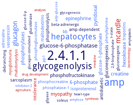
Word Map on EC 2.4.1.1

Reaction

[(1->4)-alpha-D-glucosyl]n
+
phosphate
=
[(1->4)-alpha-D-glucosyl]n-1
+
alpha-D-glucose 1-phosphate

Synonyms

1,4-alpha-glucan phosphorylase, alpha-1,4 glucan phosphorylase, alpha-1,4-glycan phosphorylase, alpha-glucan phosphorylase, alpha-glucan phosphorylase H, alpha-glucan/maltodextrin phosphorylase, alphaGP, amylopectin phosphorylase, amylophosphorylase, CcStP, cyosolic phosphorylase, GlgP, glucan phosphorylase, glucosan phosphorylase, glycogen phosphorylase, glycogen phosphorylase a, glycogen phosphorylase b, glycogen phosphorylase-a, GP, GP b, GPA, GPase, GPase a, GPb, GPBB, GPH, granulose phosphorylase, MalP, maltodextrin phosphorylase, More, muscle glycogen phosphorylase, muscle phosphorylase, muscle phosphorylase a and b, myophosphorylase, PF1535, Phb, PHO, Pho 2, Pho1, phosphorylase a, phosphorylase b, phosphorylase, alpha-glucan, plastidial phosphorylase, polyphosphorylase, potato phosphorylase, RMGPa, rmGPb, SP, starch phosphorylase, starch phosphorylase H, stGP, StP, tGPGG, type L alpha-glucan phosphorylase

ECTree

     2 Transferases
         2.4 Glycosyltransferases
             2.4.1 Hexosyltransferases
                2.4.1.1 glycogen phosphorylase

Crystallization

Crystallization on EC 2.4.1.1 - glycogen phosphorylase

Please wait a moment until all data is loaded. This message will disappear when all data is loaded.
CRYSTALLIZATION (Commentary)
ORGANISM
UNIPROT
LITERATURE
ternary complexes of maltodextrin phosphorylase with natural oligosaccharide and phosphate mimicking anions: nitrate, sulphate and vanadate. Electron density maps obtained from crystals grown in presence of Al(NO3)3 show a nitrate ion instead of the expected AlF4- in the catalytic site. The trigonal NO3- is coplanar with the Arg569 guanidinium group and mimics three of the four oxygen atoms of phosphate. The ternary complex with sulphate shows a partial occupancy of the anionic site
crystallization and structure determination of liver and muscle enzymes
dialysis against 50 mM Tris and 40 mM 2-mercaptoethanol, pH 6.8 at 0°C
-
hanging drop method, crystal structure of liver phosphorylase bound to a glycogen targeting subunit-derived peptide. C-terminus of glycogen targeting subunit of protein phosphatase 1 binds in a hydrophobically collapsed conformation to the allosteric regulator-binding site at the phosphorylase dimer interface
in complex with inhibitor (2E,2'E)-N,N'-pentane-1,5-diylbis[3-(3,4-dichlorophenyl)acrylamide]. Inhibitor is bound at the dimer interface site, the 3,4-dichlorophenyl moiety interacts hydroühobically with the enzyme
in complex with inhibitors 4-([[(2-chloro-4,5-difluorophenyl)carbonyl]carbamoyl]amino)-3-(trifluoromethoxy)benzoic acid, 1-[2-([[(2-chloro-4,5-difluorophenyl)carbonyl]carbamoyl]amino)-4-fluorophenyl]piperidine-4-carboxylic acid, and 1-(2-carboxyphenyl)-6-[(2-chloro-4,6-difluorophenyl)amino]-4-oxo-1,2,3,4-tetrahydroquinoline-3-carboxylic acid. Binding to inhibitor 4-([[(2-chloro-4,5-difluorophenyl)carbonyl]carbamoyl]amino)-3-(trifluoromethoxy)benzoic acid is exclusively enthalpic. The inhibitors 1-[2-([[(2-chloro-4,5-difluorophenyl)carbonyl]carbamoyl]amino)-4-fluorophenyl]piperidine-4-carboxylic acid, and 1-(2-carboxyphenyl)-6-[(2-chloro-4,6-difluorophenyl)amino]-4-oxo-1,2,3,4-tetrahydroquinoline-3-carboxylic acid fully exploit the volume of the binding pocket and show pronounced binding entropy
in complexwith inhibitor 5-chloro-N-[4-(1,2-dihydroxyethyl)phenyl]-1H-indole-2-carboxamide, inhibitor binds to a solvent cavity at the dimer interface, with the two hydroxyl groups making favorable electrostatic interactions with the enzyme
crystal structure of glycogen phosphorylase beta-glucopyranosylidene spirothiohydantoin complex at 2.26 A resolution
-
crystal structure of muscle glycogen phosphorylase a in complex with glucose and in complex with both glucose and CP320626 at 2.0 A resolution
-
crystal structure of the cocrystallized rabbit muscle glycogen phosphorylase b–FR258900 complex, 2.2 A resolution
determination of crystal structures of the glycogen phosphorylase b-gallic acid and glycogen phosphorylase b-ellagic acid complexes, overview
enzyme complexed with inhibitor beta-D-glucopyranosyl bismethoxyphosphoramidate by soaking of native enzyme crystals in 150 mM inhibitor 7.5 h prior to diffraction data collection, X-ray diffraction structure determination and analysis at 1.83 A resolution, modeling
enzyme in complex with different inhibitory N-(4-substituted-benzoyl)-N'-(beta-D-glucopyranosyl)urea compounds, X-ray diffraction structure determination and analysis at 1.8 A resolution, molecular modelling
-
in comlex with inhibitor N-[1-(2-amino-2-oxoethyl)-2-oxo-1,2,3,4-tetrahydroquinolin-3-yl]-2-methyl-6H-thieno[2,3-b]pyrrole-5-carboxamide, localization of two inhibitor molecules at the enzyme dimer interface
-
in complex with inhibitors (2R,3R,4S,5R,6R)-3,4,5-trihydroxy-2-hydroxymethyl-7,9-diaza-1-oxa-spiro[4,5]decane-8,10-dione, (2R,3R,4S,5R,6R)-3,4,5,9-tetrahydroxy-2-hydroxymethyl-7,9-diaza-1-oxa-spiro[4,5]decane-8,10-dione, 1-deoxy-1-methoxycarbonylamino-beta-D-glucopyranose. Inhibitors bind at the active site of the enzyme and induce minor movements of the side chains of D283 and N284 that block access of the substrate glycogen to the catalytic site
in complex with inhibitors beta-D-glucopyranosyl 1-oxalylamide, beta-D-glucopyranosyl 1-(methoxy(oxo)acetyl)-amide, beta-D-glucopyranosyl 1-(2-cyclopropylamino-2-oxoacetyl)-amide. Inhibitors accomodate at the catalytic site at approximately the position of alpha-D-glucose and stabilize the T-state conformation
-
native enzyme crystals are soaked in 150 mM gamma-cyclodextrin, or 15 mM beta-cyclodextrin, respectively, and 70 mM maltopentaose or maltoheptaose in fesh solutions of mother liquor containing 10 mM MES, pH 6.7, 0.1 mM EDTA, at least 2 h at room temperature, X-ray diffraction structure determination and analysis of enzyme-inhibitor complexes at 2.3 A and 1.94 A resolution, respectively
-
purified enzyme bound to different inhibitors, X-ray diffraction structure determination and analysis at 1.9-2.0 A resolution, structure modeling
-
purified enzyme bound to diverse inhibitor molecules, X-ray diffraction structure determination and analysis, modeling
-
purified native muscle enzyme, soaking of crystals in a 5 mM inhibitor solution in 10 mM sodium N,N-bis(2-hydroxyethyl)-2-aminoethanesulfonate, pH 6.7, containing 0.1 mM EDTA, and 0.02% w/v sodium azide at room temperature for 2 h prior to data collection, X-ray diffraction structure determination and analysis at 1.85-2.15 A resolution
hanging-drop vapor diffusion, crystals diffract to 2.4 A resolution
-