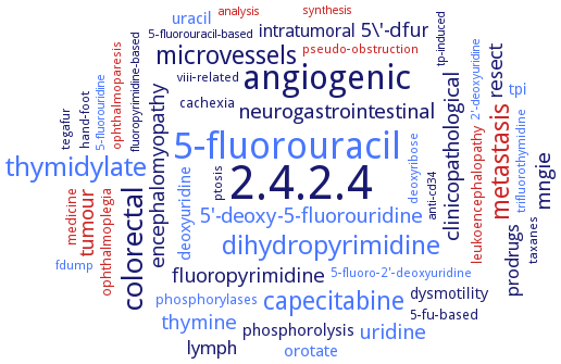
2.4.2.4: thymidine phosphorylase
This is an abbreviated version!
For detailed information about thymidine phosphorylase, go to the full flat file.

Word Map on EC 2.4.2.4 
-
2.4.2.4
-
5-fluorouracil
-
angiogenic
-
colorectal
-
thymidylate
-
dihydropyrimidine
-
capecitabine
-
microvessels
-
metastasis
-
clinicopathological
-
uridine
-
mngie
-
neurogastrointestinal
-
tumour
-
5'-deoxy-5-fluorouridine
-
fluoropyrimidine
-
thymine
-
5\'-dfur
-
resect
-
encephalomyopathy
-
prodrugs
-
lymph
-
intratumoral
-
deoxyuridine
-
uracil
-
orotate
-
phosphorolysis
-
dysmotility
-
tpi
-
cachexia
-
phosphorylases
-
leukoencephalopathy
-
ophthalmoplegia
-
medicine
-
5-fu-based
-
ptosis
-
taxanes
-
pseudo-obstruction
-
tegafur
-
deoxyribose
-
5-fluoro-2'-deoxyuridine
-
viii-related
-
hand-foot
-
fdump
-
ophthalmoparesis
-
trifluorothymidine
-
analysis
-
5-fluorouridine
-
anti-cd34
-
5-fluorouracil-based
-
tp-induced
-
2'-deoxyuridine
-
synthesis
-
fluoropyrimidine-based
- 2.4.2.4
- 5-fluorouracil
-
angiogenic
- colorectal
- thymidylate
- dihydropyrimidine
- capecitabine
-
microvessels
- metastasis
-
clinicopathological
- uridine
-
mngie
-
neurogastrointestinal
- tumour
- 5'-deoxy-5-fluorouridine
-
fluoropyrimidine
- thymine
-
5\'-dfur
-
resect
-
encephalomyopathy
-
prodrugs
- lymph
-
intratumoral
- deoxyuridine
- uracil
- orotate
-
phosphorolysis
-
dysmotility
- tpi
-
cachexia
- phosphorylases
- leukoencephalopathy
- ophthalmoplegia
- medicine
-
5-fu-based
-
ptosis
-
taxanes
- pseudo-obstruction
-
tegafur
- deoxyribose
- 5-fluoro-2'-deoxyuridine
-
viii-related
-
hand-foot
- fdump
- ophthalmoparesis
- trifluorothymidine
- analysis
- 5-fluorouridine
-
anti-cd34
-
5-fluorouracil-based
-
tp-induced
- 2'-deoxyuridine
- synthesis
-
fluoropyrimidine-based
Reaction
Synonyms
angiogenic factor platelet-derived endothelial cell growth factor/thymidine phosphorylase, animal growth regulators, blood platelet-derived endothelial cell growth factors, blood platelet-derived endothelial cell growth factor, deoA, deoxythymidine phosphorylase, dthdpase, EC 2.4.2.23, gliostatin, gliostatins, GLS, HTP, More, PD-ECGF, PD-ECGF/TP, PD-ECGF/TP platelet-derived endothelial cell growth factor, PDECGF, phosphorylase, thymidine, platelet derived endothelial cell growth factor, platelet derived endothelial cell grwoth factor, platelet derived-endothelial cell growth factor, platelet-derived endothelial cell growth factor, platelet-derived endothelial cell growth factor/thymidine phosphorylase, platelet-derived endotherial cell growth factor, ppnP, pynpase, pyrimidine deoxynucleoside phosphorylase, pyrimidine nucleoside phosphorylase, pyrimidine phosphorylase, pyrimidine/purine nucleoside phosphorylase, TDRPASE, ThdPase, thymidine phosphorylase, thymidine Pi deoxyribosyltransferase, thymidine-orthophosphate deoxyribosyltransferase, thymidine:orthophosphate deoxy-D-ribosyltransferase, Tpase, TYMP


 results (
 results (
 top
top


